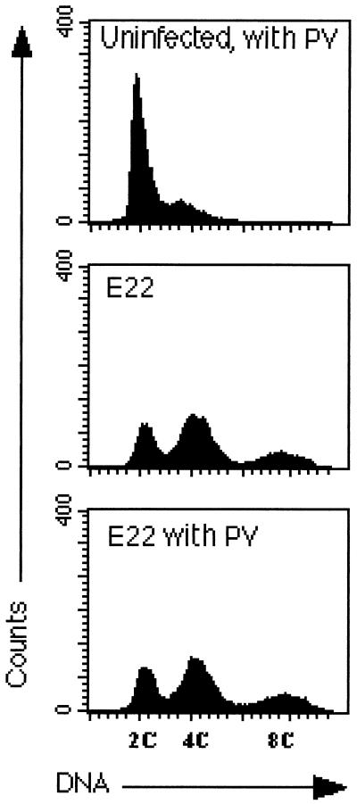
FIG. 5.
Distribution of HeLa cells according to DNA content 72 h after the interaction in the presence or absence of the tyrosine phosphatase inhibitor PV. Cells were infected with E22 for 2 h, PV was added, and the interaction was continued for 2 h. After several washes, the cells were incubated for 72 h without bacteria and PV, and then the cell distribution according to DNA content was determined by flow cytometry.

