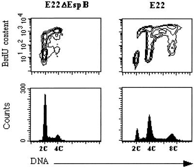
FIG. 6.
Determination of BrdU incorporation in E22ΔEspB- and E22-exposed cells by bivariate flow cytometry. Seventy-two hours after exposition to bacteria, cells were treated for 6 h with BrdU (5 μg/ml). Incorporated BrdU was labeled by FITC indirect immunofluorescence, and DNA was labeled with propidium iodide. Contour maps of DNA red fluorescence versus FITC fluorescence are shown on the upper row, and corresponding DNA frequency distributions are displayed below.

