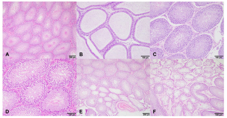
Figure 3.
Histological sections of the testis and epididymis of male rats of control group (A,D) and treated groups (B,C,E,F) at different dosages of isoflavones. (A,D): Normal seminiferous tubules and normal Leydig cells; (B): presence of a normal sperm reserve in the epididymis of a rat treated with a low doses of isoflavones at the beginning of treatment; (C): testis of a rat treated with high doses of isoflavones at the beginning of treatment, with no histopathological changes in the germinal epithelium of seminiferous tubules and presence of mature spermatozoa in the lumen; and (E,F): testicular degeneration of rats treated with low (E) and high (F) doses of isoflavones at the end of treatment. Most seminiferous tubules and less than 25% of tubules were degenerated in low-dosage- and high-dosage-treated groups, respectively. Degenerated seminiferous tubules had thickness of the basement membranes and an absence of germinal epithelium, being lined exclusively by Sertoli cells (Sertoli cell pattern).

