. 2023 Mar 10;12:e81405. doi: 10.7554/eLife.81405

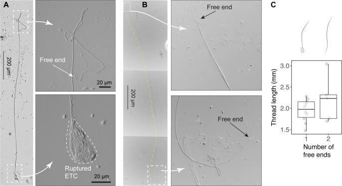
Figure 4. Size of released epidermal threads.

(A) A partially released thread (~2 mm long) from a ruptured epidermal thread cell (ETC), as viewed under light microscopy (see also Figure 4—figure supplement 1). (B) A thread with two free ends. (C) Boxplots of thread length (LT) measurements based on threads with one free end (N=10) and two free ends (N=5). Values are length from individual threads. For threads with one free end, LT = 1.95 ± 0.27 mm (mean ± SD); for threads with two free ends, LT = 2.2 ± 0.54 mm, with this mean value used in scaling models.

Figure 4—source data 1. Length of epidermal threads sampled using transmitted light microscopy.

Figure 4.

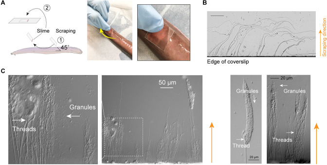
Figure 4—figure supplement 1. Release of epidermal threads.

Figure 4—figure supplement 1.

(A) Schematic of sampling epidermal mucus using a coverslip scraped along the skin of an anesthetized hagfish. The coverslip was only gently pressed against the hagfish skin, as shown on the right. (B) Epidermal threads captured by the edge of coverslips, as viewed under light microscopy. (C) After scraping hagfish skin with the edge of a coverslip, ruptured epidermal thread cells (ETCs) and free granules and threads were evident near the coverslip edge. Enlarged area shows partially unraveled threads and granules.
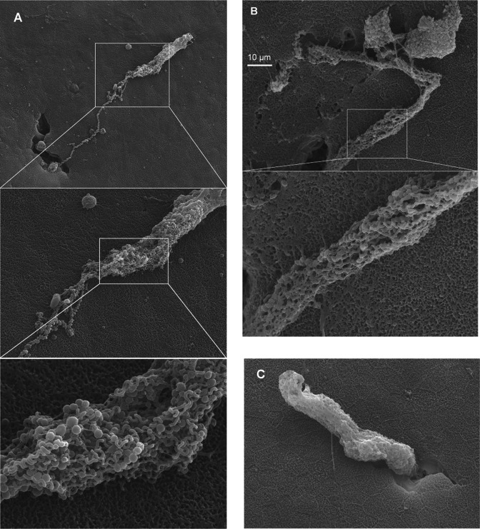
Figure 4—figure supplement 2. Release of epidermal threads.

Figure 4—figure supplement 2.

(A–B) Granule clusters found on the epidermal surface, with helical threads still attached. (C) Partially released epidermal thread cell (ETC) granule cluster found on the epidermal surface.