. 2023 Mar 10;12:e81405. doi: 10.7554/eLife.81405

Figure 7. Water content and fibrosity of hagfish epidermal slime.

(A) The relative water content of epidermal slime collected by scraping a glass coverslip over blotted skin (unswollen) and underwater (swollen). Dots represent individual samples; colors represent different animals (N=3 for each group; see Materials and methods). (B–C) A comparison of slime composition (in relative volumes) and fibrosity between epidermal and defensive slimes. Note the high water content and low fibrosity of defensive slime produced with turbulent mixing after active ejection. See Figure 7—figure supplement 1f or more information on defensive slime.

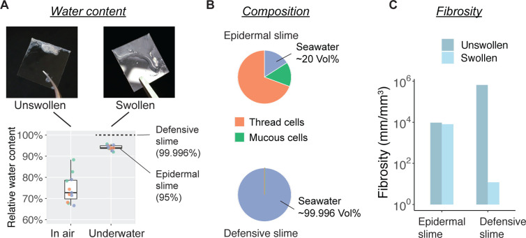
Figure 7—source data 1. Water content of epidermal slime sampled from hagfish skin (blot-dried in air versus underwater).

Figure 7.

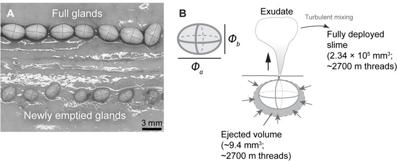
Figure 7—figure supplement 1. Morphometrics of hagfish defensive slime.

Figure 7—figure supplement 1.

(A) Full glands and newly emptied glands on the same hagfish, showing difference in size and similarity in shape (see Schorno et al., 2018). We found that full and newly emptied glands share a similar aspect ratio of ~1.44 (see Materials and methods, Section ‘Fibrosity of defensive slime’). (B) Top left shows the configuration of major and minor axes for a slime gland. The right shows the configuration for estimating the volume of ejected gland exudate. The volume of ejected exudate per gland can be approximated as the volume difference between full and newly emptied glands (see Materials and methods, Section ‘Fibrosity of defensive slime).