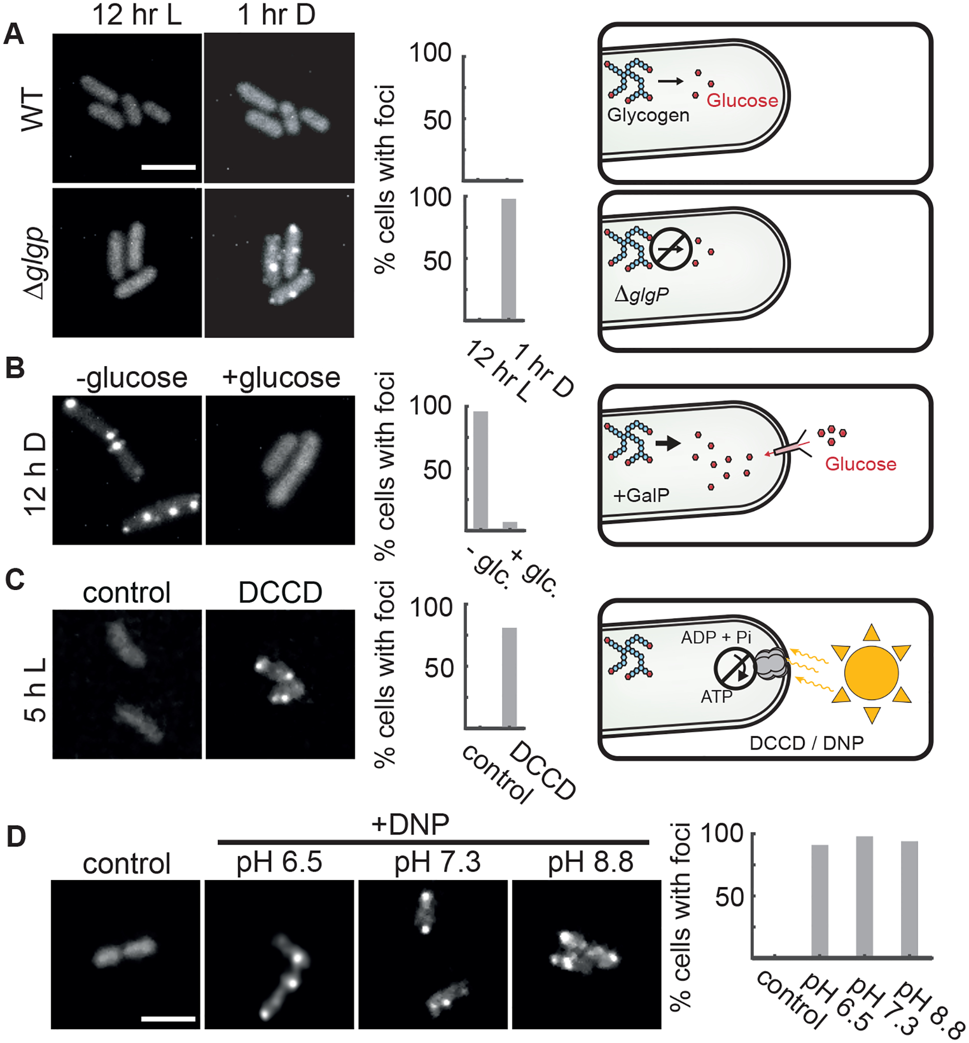
Figure 4. Metabolic Limitation Is a Key Driver of Protein Condensation.

(A) Live cell imaging of MetX-EYFP in the light (left) or after 1 h in darkness (right) in either wildtype (top) or in a glycogen breakdown-deficient mutant (ΔglgP). Scale bar: 3μm.
(B) MetX-EYFP fluorescence in a strain expressing the GalP sugar transporter and capable of glucose-dependent growth in darkness. Images were captured after 12 h of dark treatment, with or without glucose.
(C) MetX-EYFP fluorescence in cells incubated in light when ATP synthesis was blocked (DCCD, 15 μM). Control shows the effect of 0.1% methanol vehicle. Images were taken 5 h after treatment.
(D) Representative micrographs of DNP-treated MetX-EYFP cells showing fluorescent puncta in light. The pH of the media was adjusted with Bis-Tris or Tris buffer. Scale bar is 3μm. Images were taken 5–8 h after treatment. Histograms show percentages of cells with foci, N = 50–100 cells.
