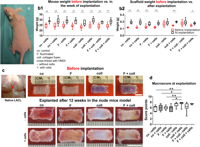
Fig. 1.
Body weight development of the animals, weight, and macroscopical appearance of the scaffold variants before implantation (after 1 week in vitro) and after explantation after 3 months in vivo. a1 Mice with a scaffold implanted into the subnuchal region. b1 Body weight of the mice in the week before scaffold implantation (red box plots) and in the week of explantation (black box plots). b2 Scaffold weight (wet weights, red box plots) pre-implantation and post explantation (black box plots). Macroscopical appearance of the lapine anterior cruciate ligament (LACL) (c1, red arrow) and the scaffold variants before implantation (c2–c5) and after explantation (c6–c13). d Results of macroscoring of the scaffolds. Co: controls; F: functionalization by gas fluorination; coll: collagen foam cross-linked with HMDI, -: implanted without cells, + : implanted with LACL-derived ligamentocytes, cultured for 1 week on the scaffold before scaffold implantation. p values: * < 0.05, ** < 0.01. Scale bars 1 cm

