Fig. 2.
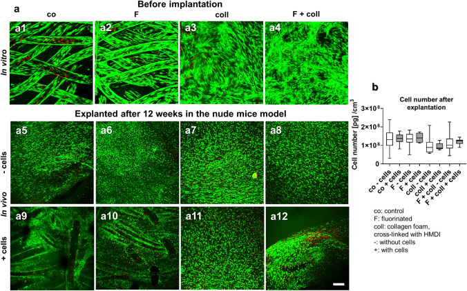
Cell viability and content in the scaffolds before implantation and after explantation after 3 months remaining in vivo. Cell viability in the scaffold variants before implantation after 1 week (a1–a4) and after explantation after 3 months in the nude mice model (a5–a12), scaffolds implanted without cells (a5–a8), and with lapine anterior cruciate ligament (LACL)-derived ligamentocytes, cultured for 1 week on the scaffold before scaffold implantation (a9–a12), control (a1, a5, a9), F (a2, a6, a10), collagen (a3, a7, a11) and F + collagen (a4, a8, a12). Living cells are stained green and dead cells are stained in red. b Measurement of DNA contents (pg DNA per cm3 scaffold) of the scaffold variants after implantation. Co: controls; F: functionalization by gas-phase fluorination; coll: collagen foam cross-linked with hexamethylene diisocyanate (HMDI), -: implanted without cells, + : implanted with LACL-derived ligamentocytes. Scale bar 100 µm

