Fig. 3.
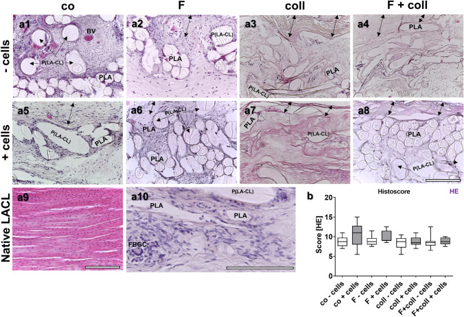
Scaffold histology after explantation after 3 months in vivo depicted by HE staining. Histology of the tissue is shown within the scaffold variants implanted without cells (-cells, a1–a4) and with lapine anterior cruciate ligament (LACL)-derived ligamentocytes (+ cells, a5–a8). Control (co, a1, a5), functionalization by gas-phase fluorination (F, a2, a6), collagen foam cross-linked with hexamethylene diisocyanate (HMDI) (coll, a3, a7) and F + coll (a4, a8). Native LACL (a9). Foreign-body giant cell and inflammatory cells visible in a co-cells scaffold variant (a10, FBGC). -cells: implanted without cells, + cells: implanted with LACL-derived ligamentocytes. BV blood vessel, PLA poly lactic acid, P(LA-CL) poly(lactic-co-ε-caprolactone). Double-headed black arrows capsule. In all images, the outer capsule surrounding the scaffold variants can be expected at the upper border. Scale bar 200 µm (a1–a9), 250 µm (a10)

