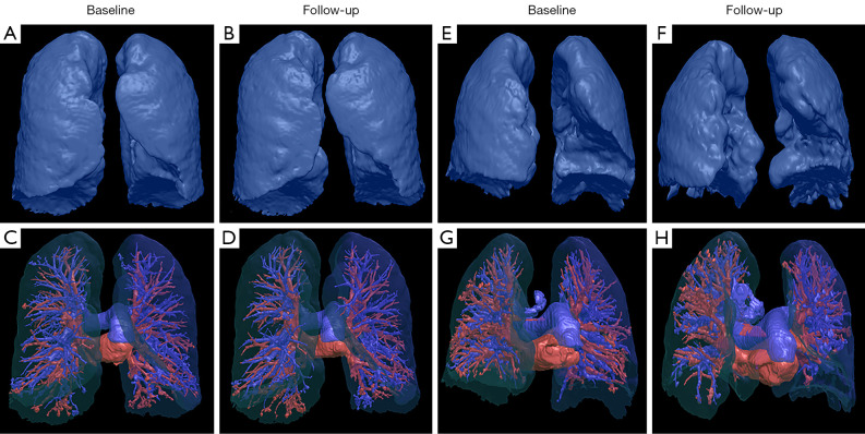
Figure 2.
Chest HRCT reconstructions of changes in vessel-related parameters during the follow-up. A 61-year-old patient diagnosed with IPF demonstrated stable disease after three years of follow-up. The follow-up HRCT showed no significant changes in lung volumes (A,B) and pulmonary vessels (C,D) compared with baseline. A 67-year-old patient diagnosed with IPF demonstrated progressive disease after two years of follow-up. The follow-up HRCT showed a significant decline in lung volume (E,F) and a significant reduced number of pulmonary vessel branches (G,H) in the predominantly left lower lung compared with baseline. HRCT, high-resolution computed tomography; IPF, idiopathic pulmonary fibrosis.

