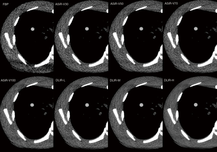
Figure 1.
Nodule images according to reconstruction method. The images were scanned at a tube current of 25 mAs and tube potential of 120 kVp. ASiR-V, adaptive statistical iterative reconstruction, DLIR, deep learning-based image reconstruction; L, low; M, medium; H, high; FBP, filtered back-projection.

