
Fig. 1. Mixed invasive ductal lobular carcinoma contains co-existing ductal and lobular histologies. a.
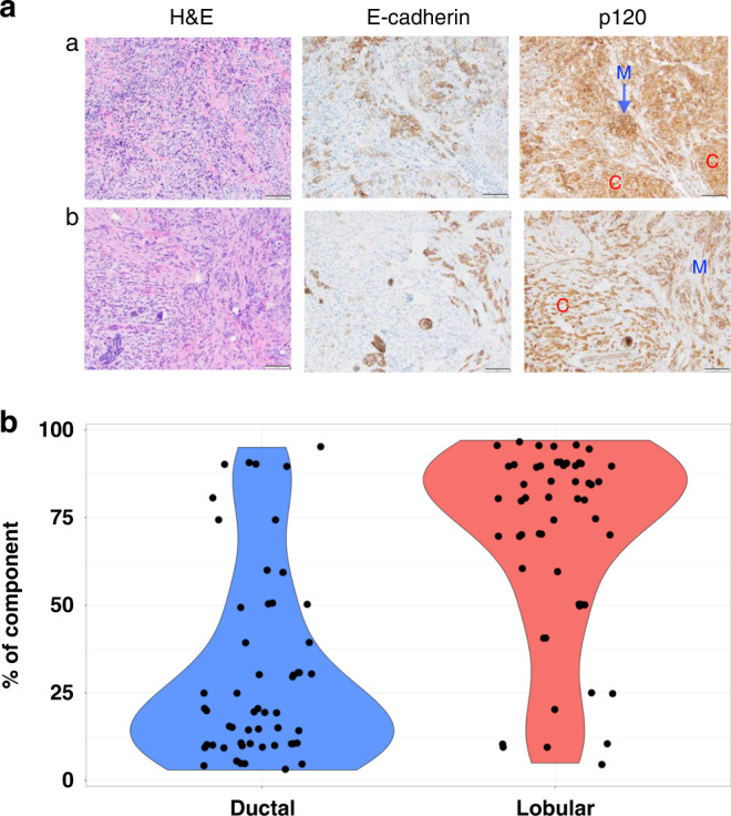
Histology examples of mixed invasive ductal lobular carcinoma. Left panel: H&E images of mDLC. Middle and right panel: IHC for E-Cadherin and b-catenin/p120. Loss of functional E-Cadherin results in the loss of lack of membranous (M) staining, and instead staining of p120 that is now accumulated in the cytoplasm (C). b Distribution of ductal and lobular components within individual mDLC tumours demonstrated by violin plot. Dots represent proportion of indicated histology within individual mDLC tumours and represent N = 54 cases. The p value from paired t-test of two components is smaller than 0.001.