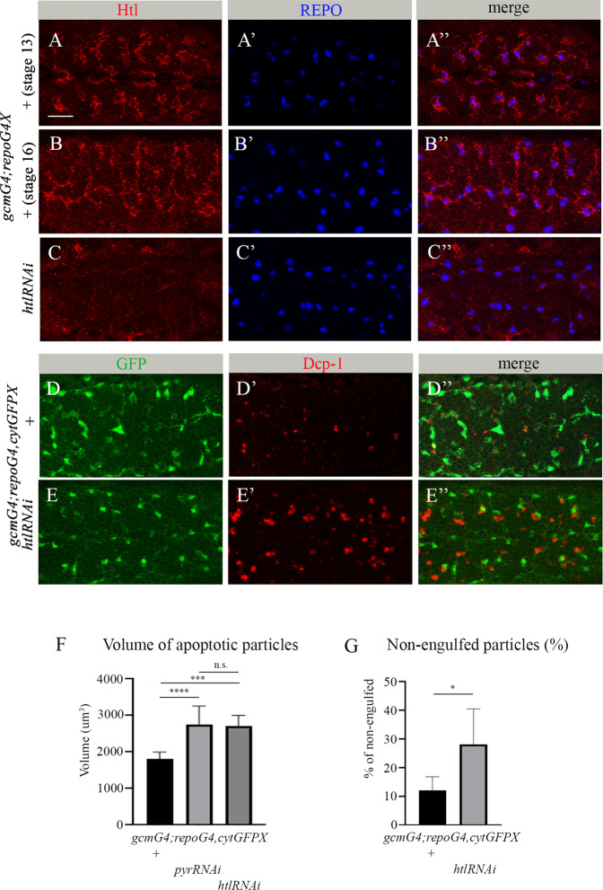
Fig. 2. htl is required in embryonic glia for phagocytosis of apoptotic neurons.
A-E” Single panes of the embryonic CNS; ventral view. Bar, 20 µm. A-B” Control embryos at stage 13 (A-A”) and stage 16 (B-B”) and (C-C”) embryos at stage 16 expressing htl RNAi in glia (gcmGal4/+;repoGal4,cytGFP/UAShtlRNAi) stained with anti-Htl (red) and anti-REPO (blue) antibodies. D-D” Stage 16 control embryos and E-E” embryos expressing htl RNAi in glia (gcmGal4/+;repoGal4,cytGFP/UAShtlRNAi) stained with anti-GFP (green) and anti-Dcp-1 (red) antibodies. F Mean total volume of apoptotic particles within stacks of the CNS and G percentages of apoptotic cell volume that is not engulfed by glia ± SEM, n = 9 (control), n = 6 (htl RNAi). Statistical significance was analyzed employing one-way ANOVA (F) and Student’s t test (G), ****p < 0.0001, ***p < 0.001, *p < 0.05, n.s. (non-significant) p > 0.05. Note the significant increase in volume of apoptotic cells and higher percentage of non-engulfed particles in embryos expressing htl RNAi in glia.

