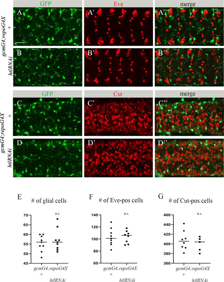
Fig. 3. Decreased htl in embryonic glia does not affect survival of embryonic neurons and glia.
A-D” Single panes of the embryonic CNS at stage 16; ventral view. Bar, 20 µm. A-A”, C-C” Control embryos (gcmGal4/+;repoGal4,cytGFP/+) and B-B”, D-D” embryos carrying glia-specific htl RNAi (gcmGal4/+;repoGal4,cytGFP/UAShtlRNAi) stained with anti-GFP (green) and anti-Eve (A’, A”, B’, B”, red) or anti-Cut (C’, C”, D’, D”, red) antibodies. Quantification of (E) REPO-positive glial cells, (F) Eve-positive neurons and (G) Cut-positive neurons in control and htl RNAi knock down embryos. Graphs represent mean total number of REPO-, Eve- or Cut-positive cells within stacks of the CNS ± SEM, n = 9 (control), n = 9, 8 and 5 (htl RNAi respectively). Statistical significance was analyzed employing Student’s t test, n.s. (non-significant) p > 0.05. Note there is no difference in neuronal and glial numbers between control and htl knock down embryos.

