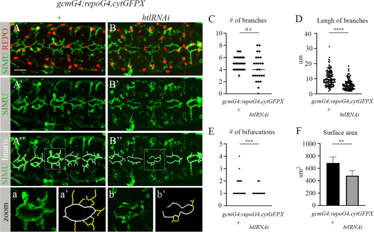
Fig. 6. Htl signaling at early embryogenesis is required for formation of the branched shape of phagocytic glia.
A-B” Single panes of the embryonic CNS at stage 16; ventral view. Bar, 20 µm. A-A”, a, a’ Control embryos (gcmGal4/UAScytGFP; +/+) and B-B”, b, b’ embryos expressing htlRNAi with gcmGal4 (gcmGal4/UAScytGFP; +/UAShtlRNAi) stained with anti-SIMU (green) and anti-Repo (red) antibodies. A”, B” Specific glial branches included in our analysis depicted with white lines using Imaris software. a, a’, b, b’ Zoom-in of rectangular areas of A” and B”, respectively. Yellow lines mark extensions protruding from the midline, evaluated and presented in graphs. Graphs represent the mean (C) number of branches; D length of branches; E number of bifurcations; and F surface area of all yellow-labeled branches ± SEM, n = 5. Statistical significance was analyzed employing one-way ANOVA, ****p < 0.0001, ***p < 0.001, **p < 0.01, n.s. (not significant) p > 0.05.

