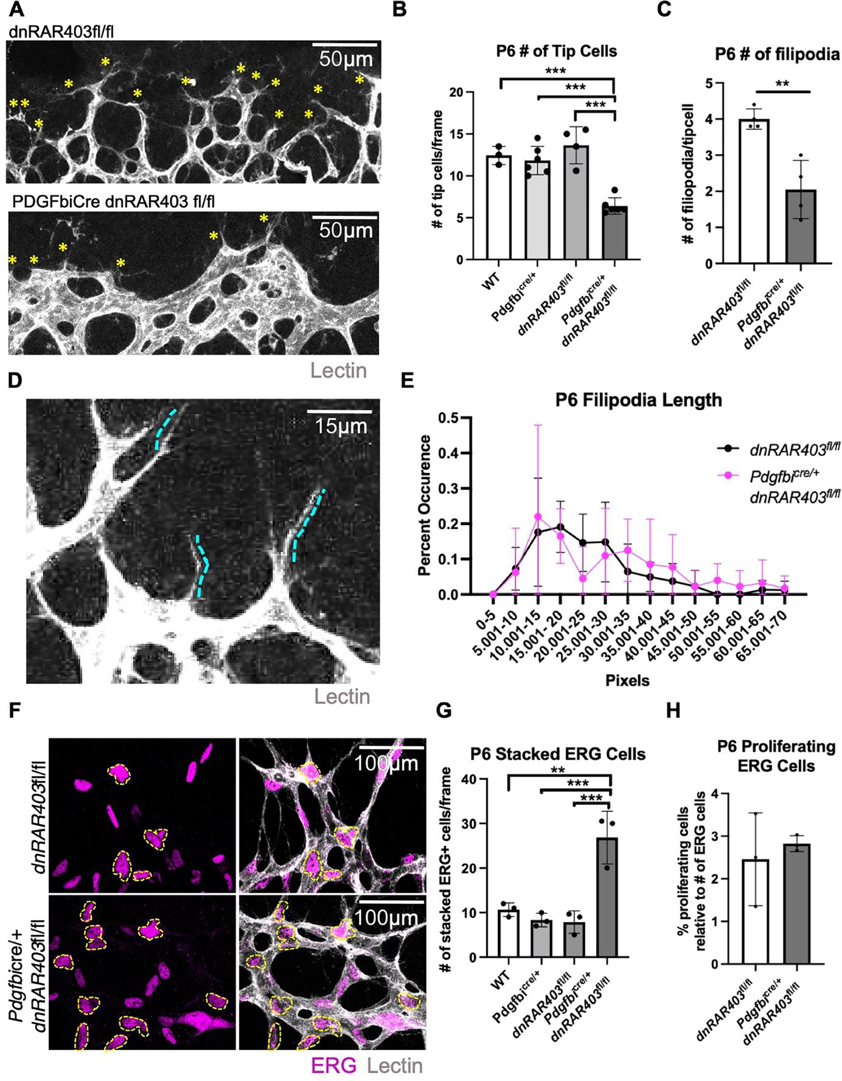
Figure 2. The loss of endothelial retinoic acid signaling reduces tip cell and filopodia number at the vascular front.

Quantification of the number of tip cells at the vascular front (A, yellow asterisks) revealed that compared to controls, Pdgfbicre/+ dnRAR403fl/fl retinas had significantly reduced tip cells (B) and the number of filipodia extended from each tip cell (C). Quantification of the length of filipodia (D) between controls and Pdgfbicre/+ dnRAR403fl/fl retinas revealed no significant difference (E). Analysis of the number of stacked endothelial cells (F, outlined in yellow) stained with ERG (magenta) and vessels stained with IB4 (white) revealed that compared to controls, Pdgfbicre/+ dnRAR403fl/fl retinas have significantly increased overlapped ERG nuclei. The percentage of proliferating endothelial cells, as labeled with EdU and ERG, were not significantly different between control and Pdgfbicre/+ dnRAR403fl/fl retinas. (p <0.05, *; p<0.01, **; p<0.001, ***).
