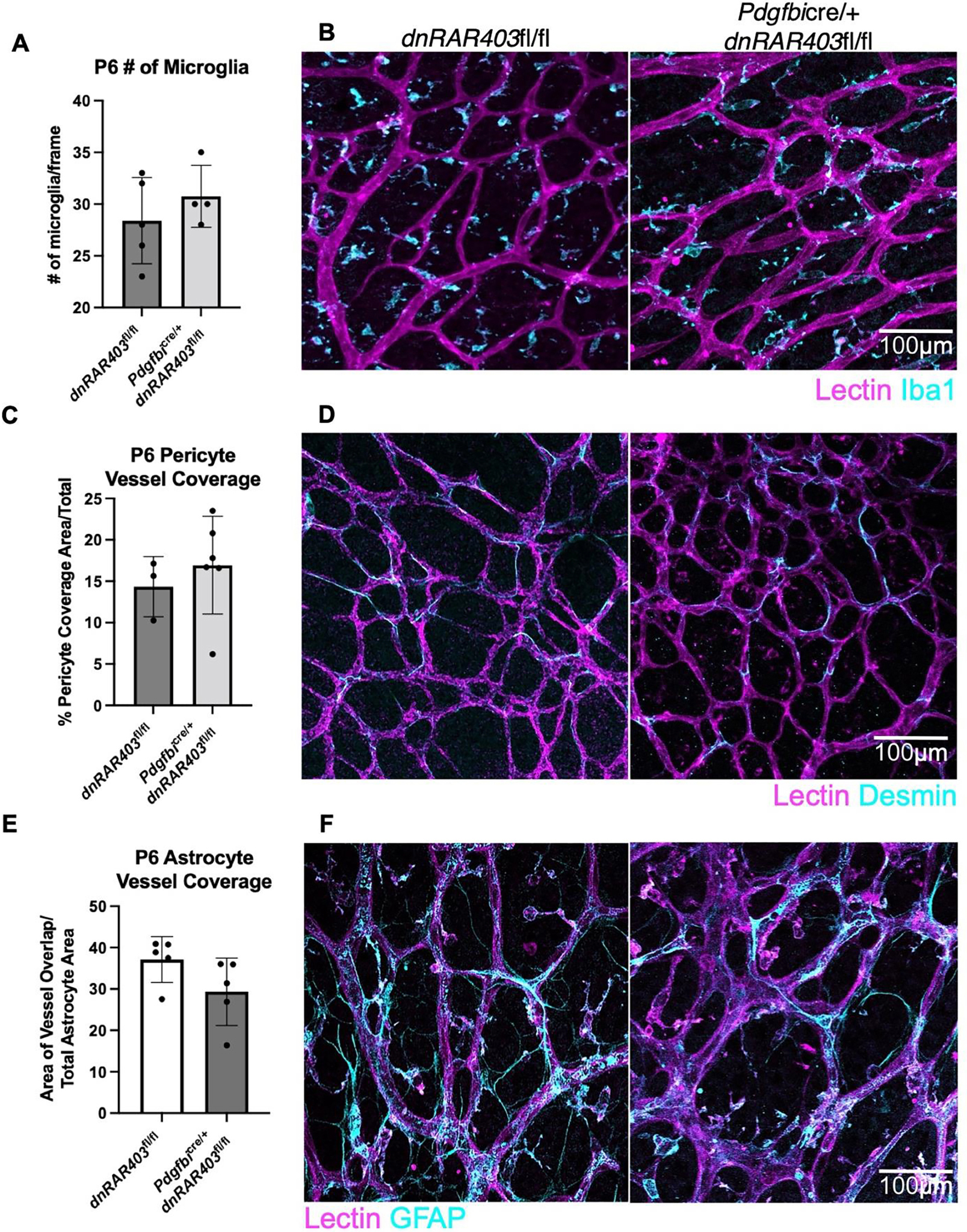
Figure 3. Microglia, pericytes and astrocytes are not overtly altered by the loss of retinoic acid signaling in endothelial cells.

Like Pdgfbicre/+ control mice, Pdgfbicre/+ dnRAR403fl/fl retinas display no quantitative difference in the number of microglia present (A) or the morphology (B, lectin, magenta; Iba1, cyan). The area of pericyte coverage of blood vessels was not significantly different between controls and Pdgfbicre/+ dnRARfl/fl retinas (C) or in morphology (D, lectin, magenta; desmin, cyan). The area of astrocyte vessel coverage was not significantly different between controls and Pdgfbicre/+ retinas (E) or in morphology (F, lectin, magenta; GFAP, cyan). (p <0.05, *; p<0.01, **; p<0.001, ***).
