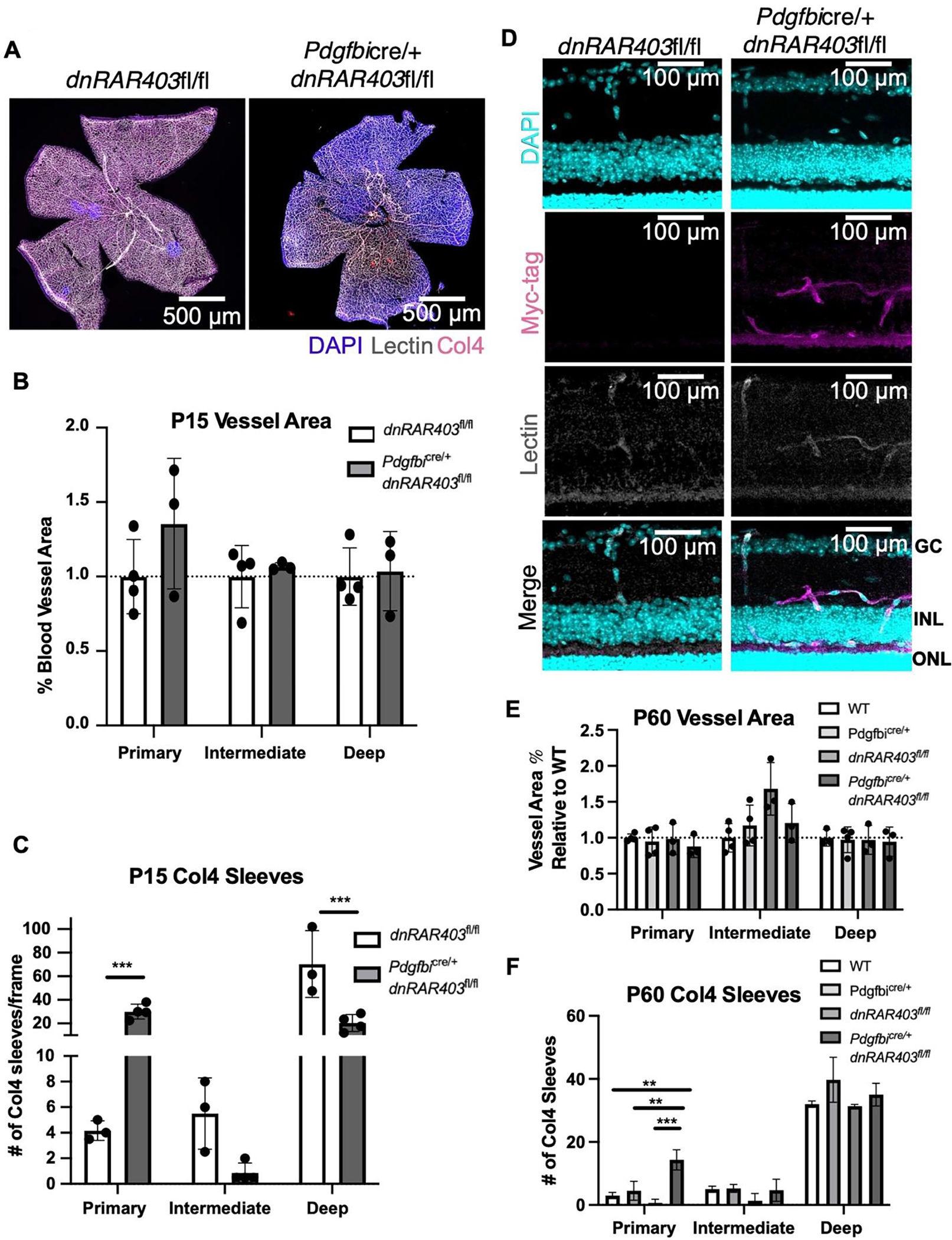
Figure 4. Angiogenesis recovers in Pdgfbicre/+ dnRAR403fl/fl retinas but there are defects in collagen 4 sleeves.

Like controls, Pdgfbicre/+ dnRAR403fl/fl retinas are fully vascularized (A) with no significant difference in vessel area (B). Analysis of Collagen-4 sleeves at P15 revealed that there were significantly more Collagen-4 sleeves in the primary plexus, no difference in the intermediate plexus, and significantly decreased sleeves in the deep plexus (C). At P60, Myc signal is observed in Pdgfbicre/+ dnRAR403fl/fl lectin+ endothelial cells in the intermediate and deep plexus (D). The blood vessel area was not significantly different from controls in the primary, intermediate, or deep plexus (E). Compared to controls, Pdgfbicre/+ dnRAR403fl/fl retinas had significantly increased Collagen-4 sleeves in the primary plexus, but no significant difference in the intermediate or deep plexus (F).
