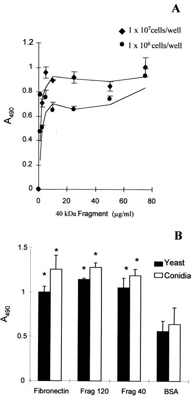
FIG. 2.
Adhesion of S. schenckii to Fn fragments. (A) Adhesion to the 40-kDa Fn fragment. Yeast cells were assayed at 107 and 106 cells/well. (B) Microtiter plates were coated with either Fn or Fn fragments: frag120 (containing the cell-binding domain) or frag40 (containing the GAG-binding domain). Control wells contained immobilized BSA only. Both conidia (open bars) and yeast cells (black bars) of S. schenckii were tested at a concentration of 107 cells per well. As described in the text, the number of bound cells was determined by measuring the optical density at 490 nm. The values are the means ± SD of triplicate wells, and this figure is representative of three independent experiments. ∗, P < 0.05.

