
Figure 2.
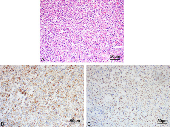
Hematoxylin and eosin staining at a medium high (×20) magnification. A: Hematoxylin and eosin staining showing the tumor tissue diffusely arranged and the ovoid, slightly larger than normal plasma cells with eccentric nuclei, coarse chromatin, and basophilic cytoplasm arranged in a spoke-wheel pattern, along with binuclear cells. B, C: Immunohistochemical staining showing MUM1(+) and CD138(+).