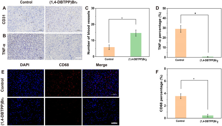
Figure 9.
Immunohistochemical staining of (A) CD31 and (B) tumor necrosis factor (TNF)-α. Quantification of (C) blood vessels and (D) inflammatory area based on CD31 and TNF-α staining, respectively. Scale bar: 200 μm. (E) Immunofluorescence staining and (F) quantification percentage of CD68 cells on day 12. Scale bar: 50 μm. *p < 0.05.

