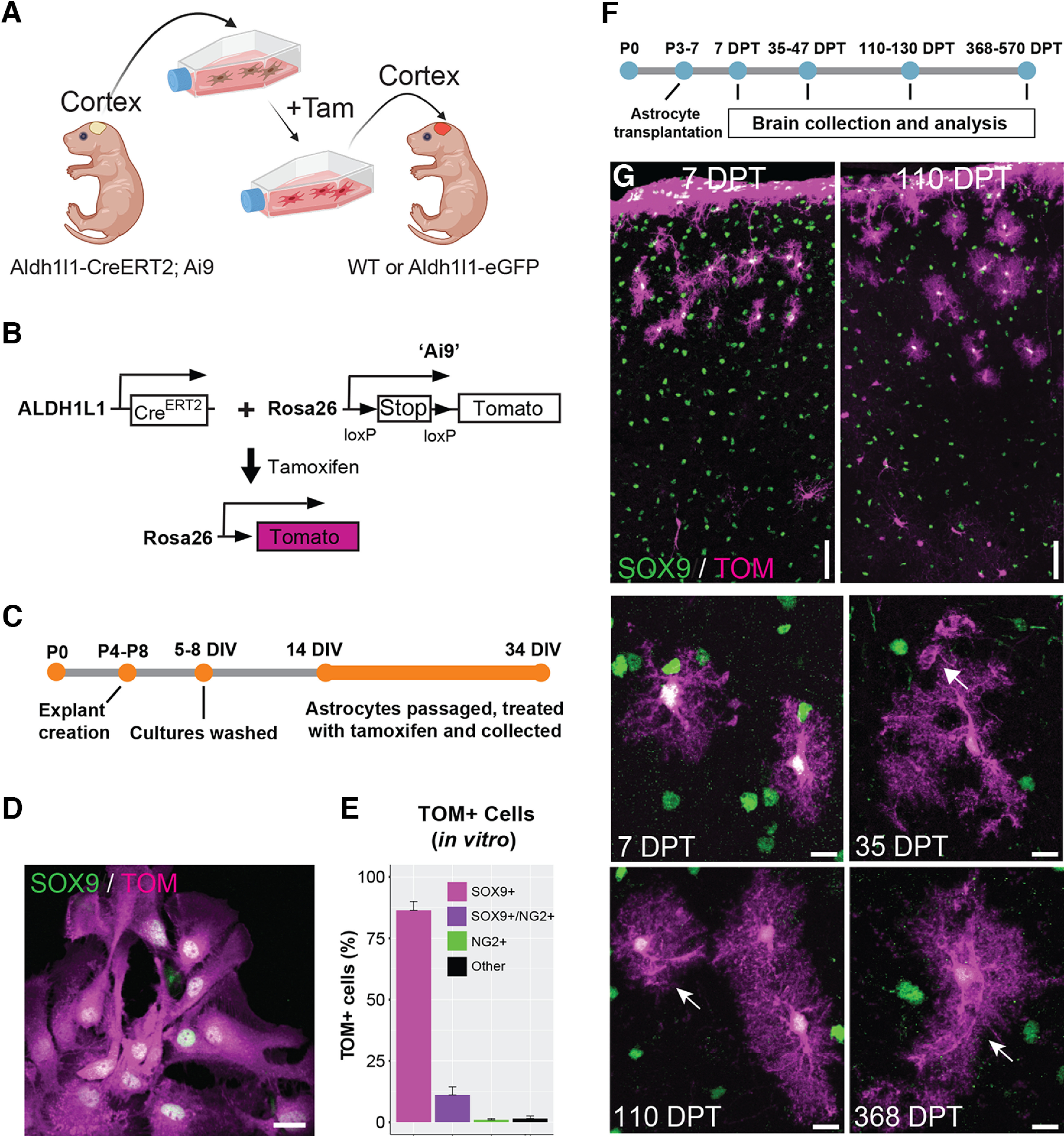
Figure 1.

Cortical astrocytes transplanted into the immature cortex display protoplasmic astrocyte morphology starting within a few days after transplantation. A, Overview of the experimental design for transplantation of cortical astrocytes into the cerebral cortex of mouse pups. Tam = 4-hydroxytamoxifen. Figure created with BioRender. B, Schematic showing the genetic background of the mice used as transplant donors. Tamoxifen = 4-hydroxytamoxifen. C, Timeline of culture preparation. D, Cultured astrocytes expressing Tomato following tamoxifen administration. Tom = Tomato. E, Percentage of Tomato+ cells in vitro expressing Sox9 (astrocytes), Sox9 and NG2 (intermediate astrocytes), NG2 (OPCs), or neither marker (“Other”). F, Timeline of astrocyte transplantation. G, Transplanted astrocytes at different days post-transplantation (DPT). Transplanted cells are labeled with Tomato (TOM) and astrocytes are detected by SOX9 immunolabeling (green). Arrows indicate astrocyte endfeet. 35 DPT = 35–47 DPT, 110 DPT = 110–130 DPT. Scale bar: 20 µm (D) and 50 and 10 µm (G, top panels, and G, middle and bottom panels, respectively).
