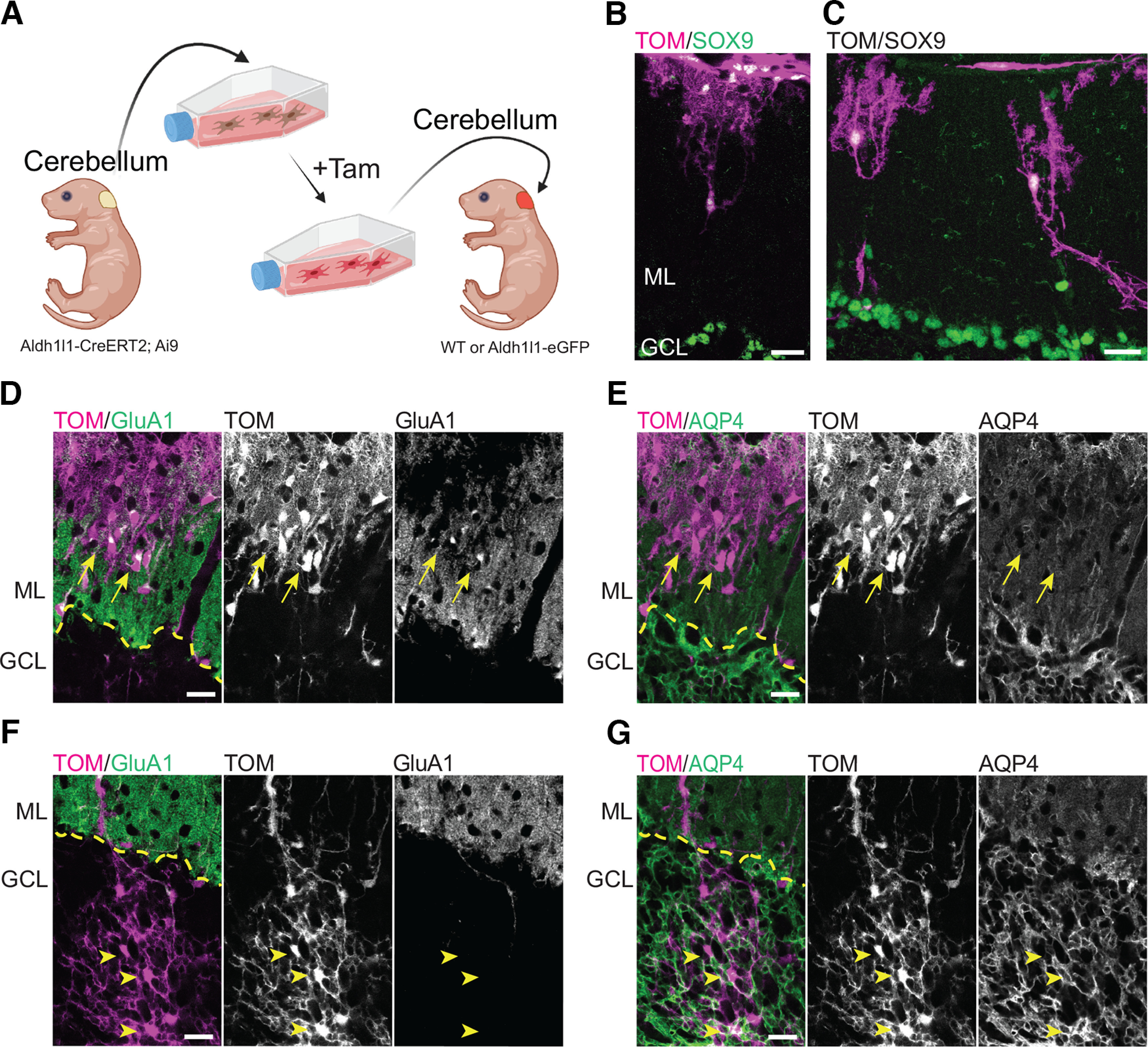
Figure 11.

Cerebellar astrocytes develop into Bergmann glia-like and velate-like astrocytes when transplanted into the cerebellum. A, Overview of the experimental design for transplantation of cerebellar astrocytes into the cerebellum of mouse pups. Tam = 4-hydroxytamoxifen. Figure created with BioRender. B–G, Cerebellar astrocytes transplanted in the cerebellar cortex acquired region-specific structural and molecular features. B, C, Transplanted cerebellar astrocytes located in the ML display BG-like morphologies. D, E, Astrocytes transplanted in the ML (arrows) express GluA1 and low levels of AQP4, like BG cells. F, G, Astrocytes transplanted in the granule cell layer (GCL; arrowheads) display a velate-like astrocytic morphology and, like velate astrocytes, are negative for GluA1 and express high levels of AQP4. Tom = Tomato. Scale bar: 20 µm (B–G). Dashed yellow line marks the interface between ML and GCL.
