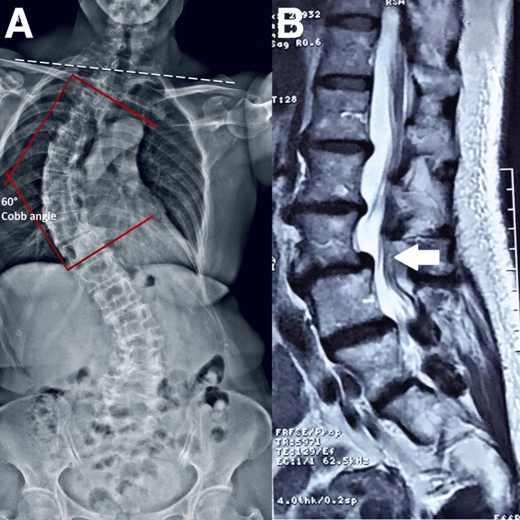
Figure 1. Full-spine radiograph and lumbar magnetic resonance image .
A) Full-spine radiograph revealing degenerative changes in the thoracic and lumbar regions, shoulder and pelvic imbalance (dash white line), and abnormal thoracic and lumbar scoliotic curves. The Cobb angle was measured at 60 ° (red lines) B) At L4-5 (white arrows), there are spondylolisthesis, posterior bulging disc, and marked bilateral facet joint degeneration, in the right facet joint and ligamentum flavum hypertrophy causing mild indentation of the thecal sac. Diffuse bulging disc and osteophytes at L2-3 causing mild indentation of the thecal sac, mild right lateral recess and mild bilateral foraminal stenosis. No nerve root compression is seen.

