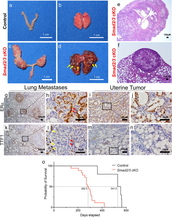
Fig. 2. Conditional deletion of SMAD2/3 results in metastatic endometrial tumors and death.
a, b Gross uterus of control (a) and Smad2/3 cKO (b) mice at 9 months of age. Smad2/3 cKO mice showed the presence of uterine masses. c, d Lungs dissected from control (c) and Smad2/3 cKO mice (d), showing that the lungs of the Smad2/3 cKO mice developed metastatic nodules (yellow arrows). e, f Cross-sections of the uterine tumor (e) and metastatic nodules (f) from Smad2/3 cKO mice stained with Hematoxylin and Eosin (H&E). g–j Immunohistochemistry of ERα in the lung nodules (g, h) or uterine tumors (i, j) from Smad2/3 cKO mice. Expression of ERα is observed in the uterine tumors (i, j), and in the lung nodules (outlined by white dotted circles), but not in the adjacent normal tumor tissue. k–n Immunohistochemistry of the lung cell marker, TTF1, in lung (k, l) and uterine tumor cross-sections (m, n) showing that neither the uterine tumors (m, n) nor metastatic nodules (k, l) express TTF1. However, the normal lung cells adjacent to the lung nodules do express TTF1 (l, yellow arrows). Red arrows in (h, l) show ERα positive cells in the lung nodules (h) that are TTF-negative in a sequential section (l). o Survival analysis comparing the survival of control mice (50% survival, 541.5 days) to Smad2/3 cKO mice (50% survival, 282.5 days).

