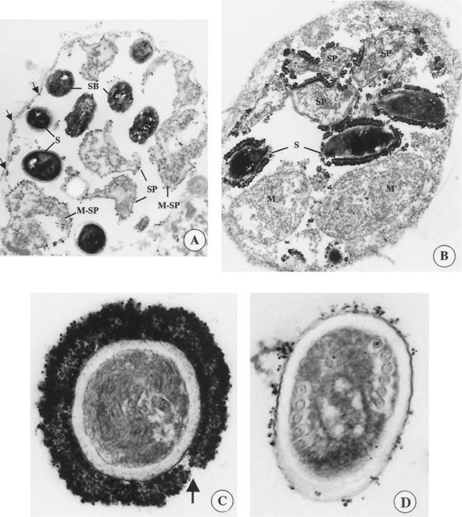
FIG. 2.
IEM of E. intestinalis-infected host cells at different developmental stages using either MAb 11B2 or 7G7 followed by a fluoronanogold anti-mouse antibody and silver-staining enhancement. (A) PV reacted with MAb 11B2. The arrows indicate residual staining along the inside of the PV lining. (B) PV reacted with MAb 7G7. (C) Cross sections of mature spores that were released from the PV reacted with MAb 7G7. The arrow indicates a gap in the exospore staining. (D) Cross sections of mature spores that have been released from the PV reacted with MAb 11B2. PVs contain cells at different stages of development: meronts (M), sporoblasts (SB), sporonts (SP), and mature spores (S). Also shown are cells that do not have a completely defined dense membrane and are considered to be in transition from meronts into sporonts (M-SP).

