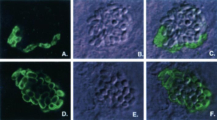
FIG. 3.
Immunofluorescence and confocal imagery of in vitro-infected host cells using MAbs specific for SWP1 (11B2) and SWP2 (7G7). (A, B, and C) Localization of SWP1. (A) Immunofluorescent staining using the SWP1-specific MAb 11B2. (B) Differential interference contrast image (Nomarski) of the same microscopic field. (C) Layering of images in panels A and B. ( D, E, and F) Localization of SWP2. (D) Immunofluorescent staining using the SWP2 MAb 7G7. (E) Differential interference contrast image (Nomarski) of the same microscopic field. (F) Layering of images in panels D and E. All images are about 16 μm wide.

