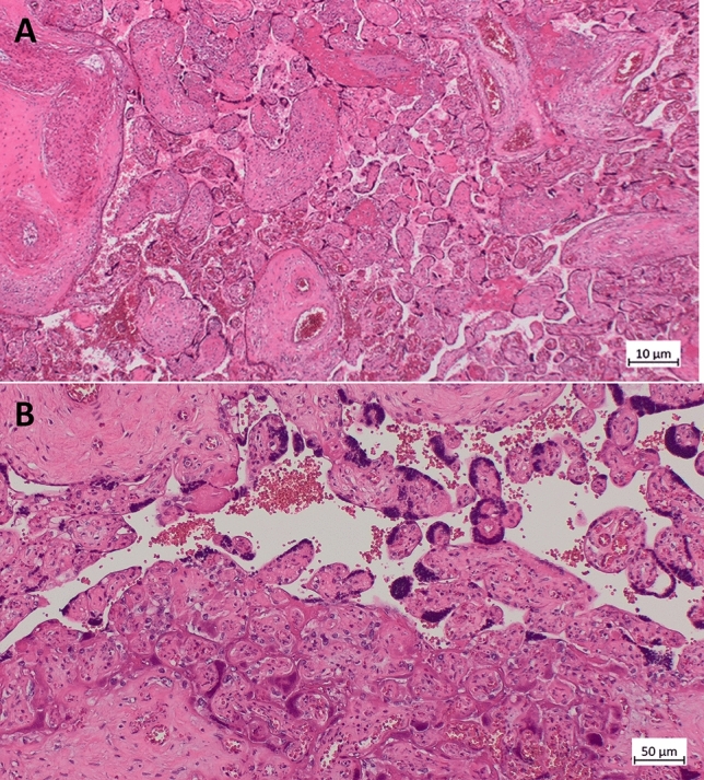
Fig. 2.

Histology (microscopic) of the placenta from Case 2: A H&E section of a placenta demonstrating necrotic syncytiotrophoblast collapse and fibrin. (200×). B collapse of the intervillous space and trophoblast necrosis (lower part), on the same image vital functioning villi (upper part) (200×)
