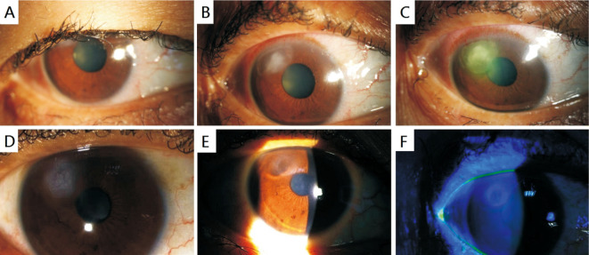
Figure 1. The slit-lamp examination.
A: There was mild conjunctival hyperaemia and eyelash trichomegaly; B: At 11 o'clock, there was a corneal infiltration and epithelial defect; C: After 5d treatment without topical sterioid application, the corneal ulcer was expanded; D: During treatment, detached eyelashes are sometimes observed adhering to corneal ulcer; E: After bandage contact lens and topical steroid, the corneal epithelium regenerated and the defect became scarred; F: The corneal epithelium was not stained with fluorescein.

