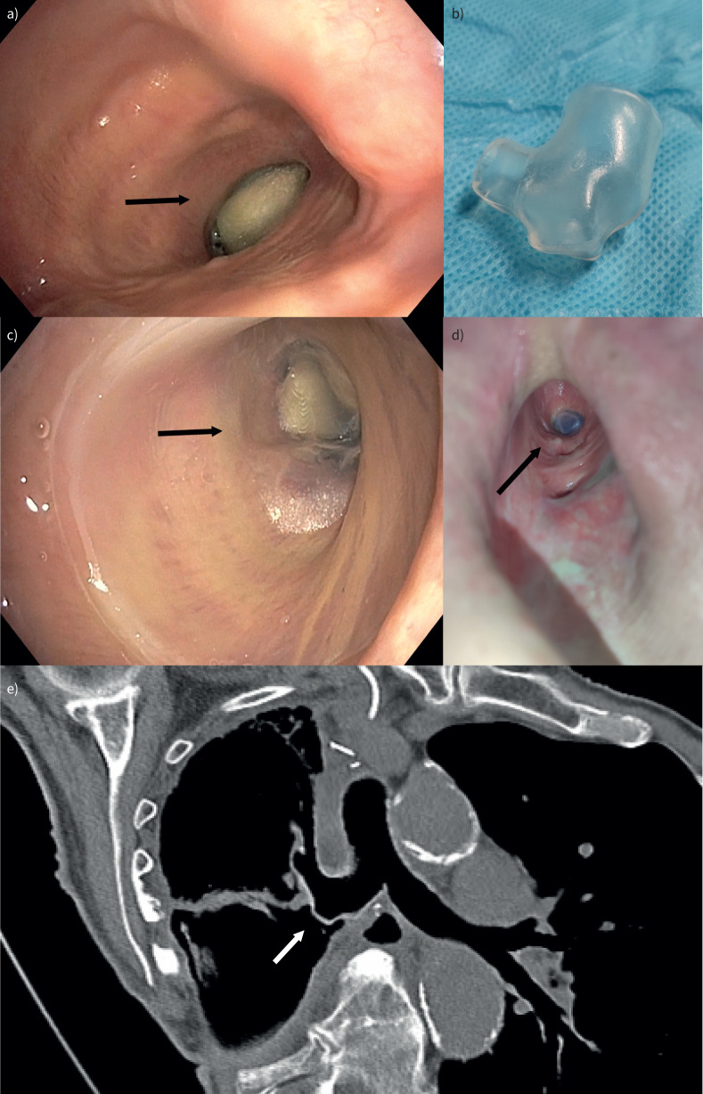
FIGURE 1.
a) Endobronchial view of the fistula before stent insertion, showing a gauze in the opening. b) Customized silicone stent using 3D reconstruction of a computed tomography scan. c) Endobronchial and d) thoracostomy views of the fistula after stent insertion, covering the fistula without occluding the right upper lobe. e) Computed tomography image showing perfect fit of the customised stent, covering the fistula.

