
Fig.4: Effects of PGE2 receptor stimulation on electrophysiological properties of DS D1-SPNs and D2-SPNs neurons.
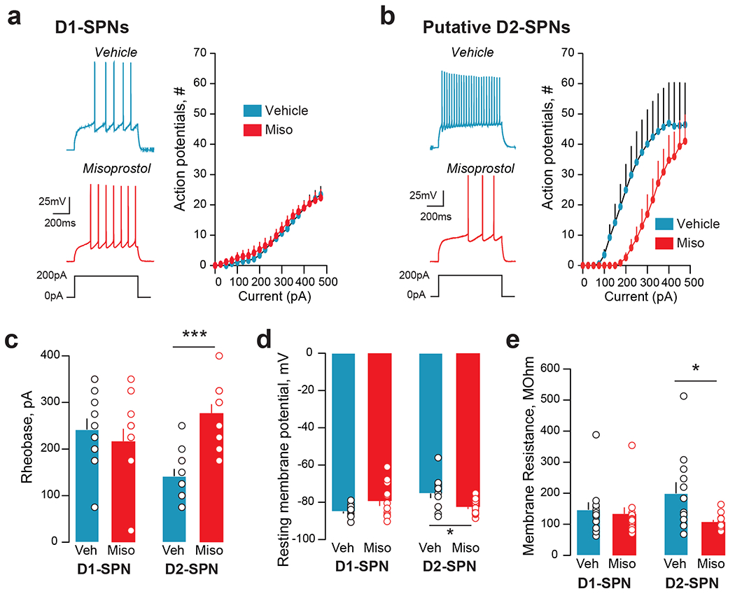
Male Drd1-Cre x Ai14 tdTomato reporter mice were injected i.p. with vehicle or misoprostol (0.1 mg.kg−1). Thirty minutes later mice were sacrificed, and brain slices were made for patch clamp electrophysiological experiments. D1- and putative D2-SPNs in the dorsomedial striatum were identified based on red fluorescence and morphology and patched. a. In current clamp, incrementally increasing depolarizing currents were injected into the cell, while action potential output was monitored. In D1-SPNs no differences occurred between cells from animals pretreated with vehicle (Veh, ncells=12; nmice=5) or with misoprostol (Miso, ncells=12; nmice=6). Left: representative examples of action potential profiles in response to a depolarizing current injection of 200 pA. Right: Average current-action potential number relationship across cells from the vehicle or misoprostol condition. Two-way repeated measures-ANOVA (RM-ANOVA), misoprostol effect not significant. b. In D2-SPNs misoprostol pre-treatment (ncells=12; mmice=5) compared to vehicle (ncells=12; nmice=5), resulted in a reduction of action potential output (RM-ANOVA, misoprostol effect, p=0.04). c. The rheobase (i.e., the minimal injected current into a neuron required to make it fire an action potential) was not affected by misoprostol pretreatment in D1-SPNs, but was significantly increased by it in D2-SPNs (2-way ANOVA, interaction, p=0.001). d. The resting membrane potential was unaltered by misoprostol in D1-SPNs, but reduced in D2-SPNs (2-way ANOVA interaction, p=0.002). e. Misoprostol reduced the membrane resistance of D2-SPNs (2-way ANOVA misoprostol effect, p=0.037). c-e, multiple comparisons with Holm-Sidak’s test, *p<0.05, ***p<0.001. See Supplementary Table 19 for detailed statistical results.