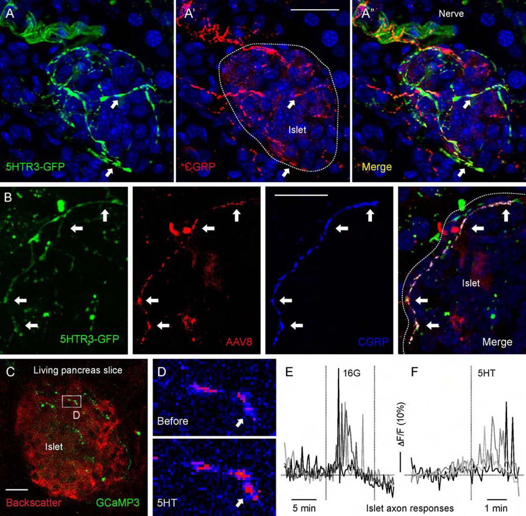
Figure 6.
Serotonin activates axonal terminals in the mouse pancreatic islet. (A-A”) Pancreatic section from a 5HT3r-GFP reporter mouse immunostained for GFP (green) and CGRP (red). (B) Pancreatic section from a 5HT3r-GFP mouse with fibers anterogradely traced by injecting AAV8-mCherry into the nodose ganglion, immunostained for GFP (green), RFP (red), and CGRP (blue). (C) z-stack of confocal images of a living pancreatic slice from a Pirt-GCaMP3 mouse. Sensory fibers (green) can be seen in an islet visualized by backscatter (red). (D) Sequential images of sensory fiber shown in B displaying a Ca2+response to serotonin (5HT, 50 μM) perfused over the slice. Arrow points to a region showing an increase in GCaMP3 fluorescence (from blue to red in pseudocolor scale). (E and F) Representative traces of mean fluorescence intensity changes in axonal terminals inside the pancreatic islet demonstrating that axonal terminals respond to an increase in glucose concentration (from 3 mM to 16 mM) and to 5HT (50 μM) stimulation. Scale bars, 20 μm.

