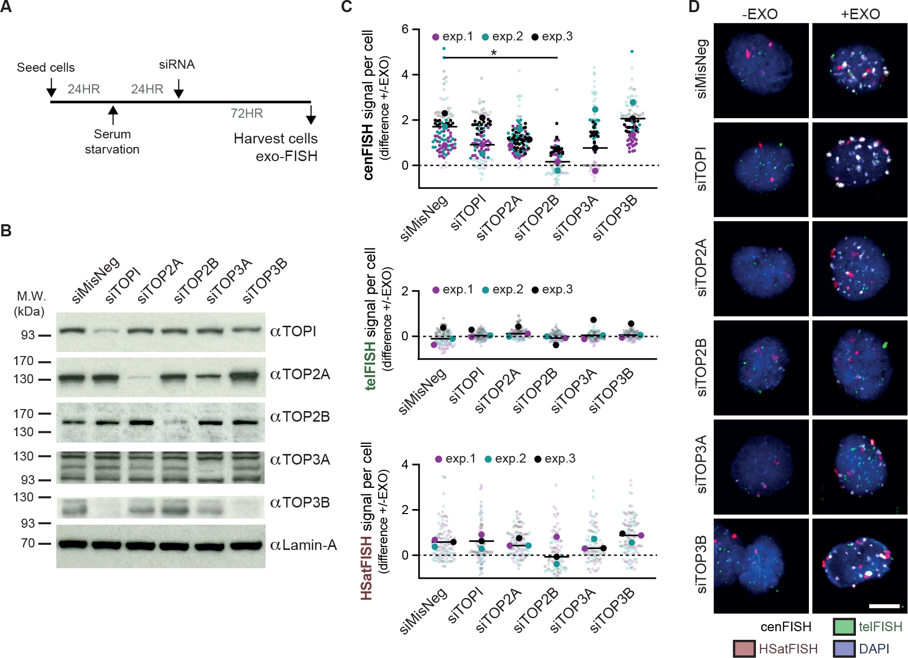
Figure 4 |. Topoisomerase IIβ induces centromere DNA breaks in quiescent cells.

(A) Experimental schematic for the depletion of the five human nuclear topoisomerases in serum-starved hTERT-RPE1 cells. hTERT-RPE1 cells were serum-starved for 24 hours prior to RNAi treatment and harvested after another 72 hours. (B) Western blot verifying the depletion of topoisomerase I (TOPI), topoisomerase IIα (TOP2A), topoisomerase IIβ (TOP2B), topoisomerase IIIα (TOP3A) and topoisomerase IIIβ (TOP3B) 72 hours after RNAi treatment with 50 nM siRNA. Lamin-A levels were used as a loading control. (C, D) Quantification and representative images of exo-FISH following 72 hours depletion of human topoisomerases. Quantification was performed as in Figure 2. The difference in exo-FISH signals between +ExoIII and −ExoIII was then calculated and plotted. Scale bar represents 10 μm. FISH signal intensity is X10,000 arbitrary units (A.U.). At least 30 cells were imaged per experimental condition. The medians of each experimental condition were used to perform a two-sided unpaired t-test (*p<0.05, **p<0.01). See also Figure S5.
