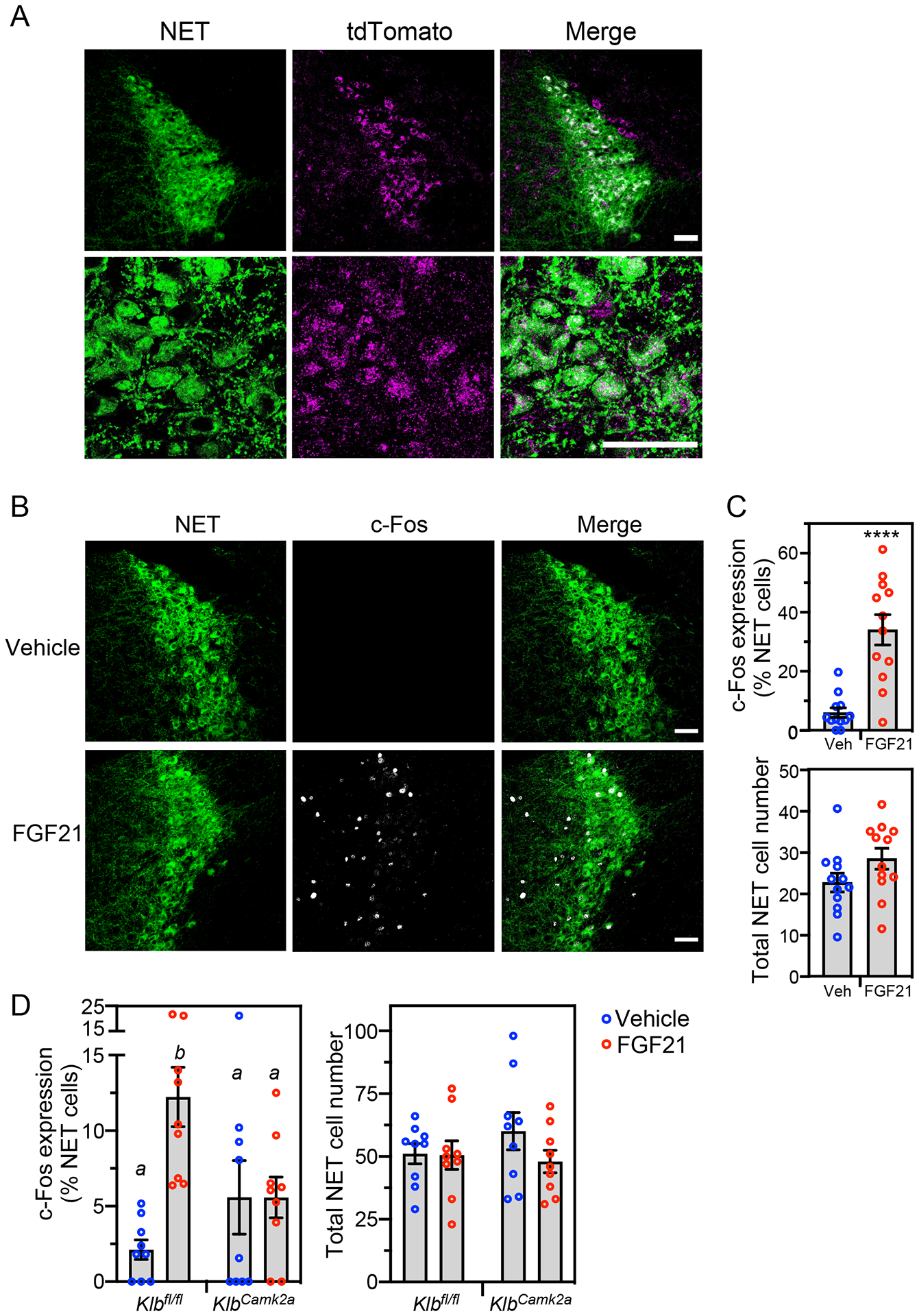
Figure 5. Pharmacologic FGF21 activates noradrenergic neurons in the locus coeruleus.

(A) Confocal images of immunostaining performed in locus coeruleus (LC) sections prepared from transgenic mice expressing tdTomato fused to the C-terminus of KLB. Antibodies against tdTomato (magenta) and norepinephrine transporter (NET; green) were used. Scale bars represent 50 μM.
(B and C) Immunostaining for c-Fos (white) and NET (green) in LC sections prepared from wild-type mice treated for 2 hours with vehicle or FGF21 (1 mg/kg, i.p.). Representative confocal images are shown in (B). Scale bars represent 50 μM. Quantification of c-Fos/NET co-expression (top panel) and total NET-positive cell number (lower panel) is shown in (C) (n = 4 sections/mouse, 3 mice/group). Data represent the mean ± SEM. ****, P < 0.0001 by Student’s t-test.
(D) Immunostaining for c-Fos and NET in LC sections from groups of control (Klbfl/fl) and neuron-specific Klb−/− (KlbCamk2a) mice treated for 2 hours with vehicle or FGF21 as in (B). Quantification of c-Fos/NET co-expression (left panel) and total NET-positive cell number (right panel) is shown (n = 3 sections/mouse, 3 mice/group). Data represent the mean ± SEM. Different lowercase letters indicate statistical significance (P < 0.05) by two-way ANOVA.
See also Figure S1.
