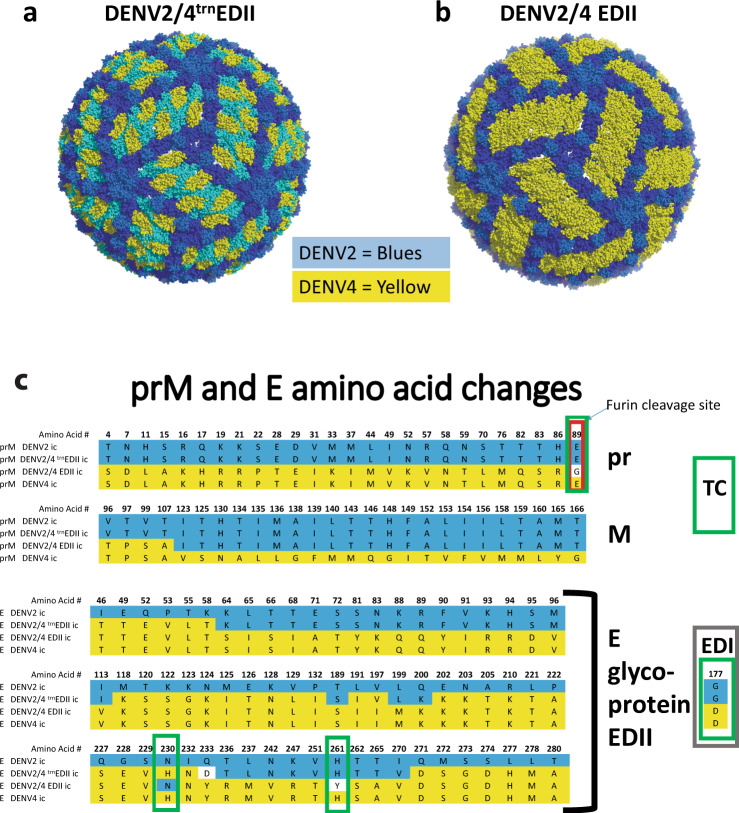
Fig. 1. Molecular models of Virions.
a PyMOL software-generated representations of changed residues. DENV2/4trnEDII virion with a truncated EDII transplanted DENV4 region in yellow. DENV2 backbone in shades of blue, EDI - dark blue, EDII – light blue, EDIII – medium blue. b DENV2/EDII virion with transplanted DENV4 EDII in yellow. DENV2 backbone in shades of blue, EDI - dark blue, EDIII – medium blue. c Amino acid changes to prM and E glycoproteins. Chimeric prM is shown in comparison with parental DENV2 and DENV4 prMs. In the chimeric DENV2/4 prM, amino acids 1-107 are from DENV4 and amino acids 108-166 are from DENV2 with the exception of E89G, which is an essential tissue culture adaptation found in recovered DENV2/4EDII. Chimeric E glycoproteins from DENV2/4trnEDII and DENV2/4EDII with their respective tissue culture mutations are shown in comparison with parental DENV2 and DENV4 sequences. DENV2 amino acids are shown in blue and DENV4 are yellow. Amino acids unique to the chimeras have white background. All amino acids not shown are homologous to DENV2 and DENV4. The furin cleavage site in prM is denoted by the red box. Tissue culture (TC) changes are denoted by green boxes. The single amino acid change in EDI is shown in a brown box.

