Fig. 5.
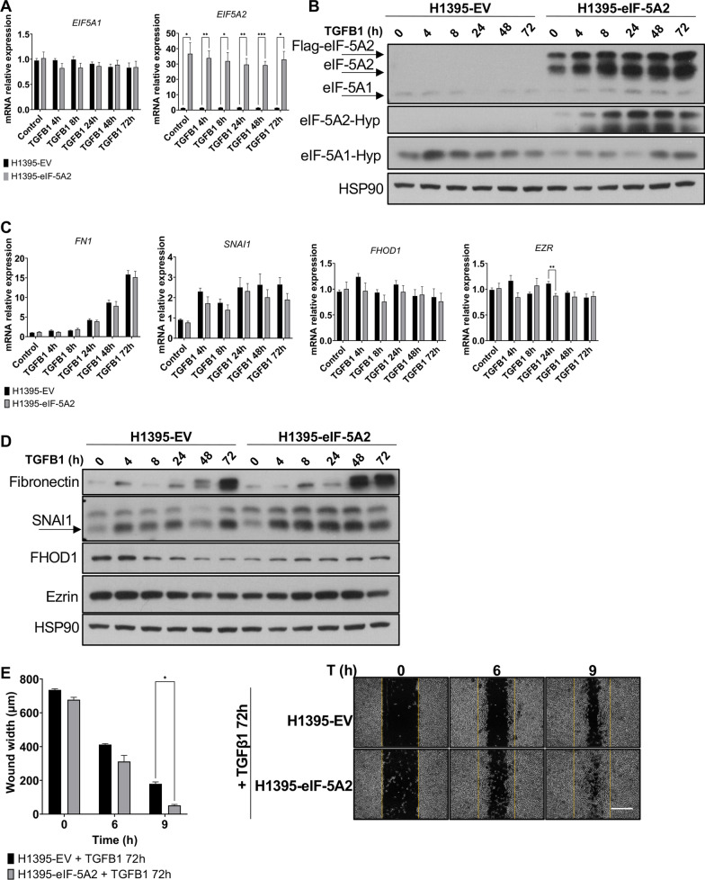
TGFB1 induces eIF-5A2 hypusination and eIF-5A2-dependent expression of EMT proteins. A EIF5A1 and EIF5A2 mRNA levels in H1395-EV and H1395-eIF-5A2 cells treated with TGFB1 for the indicated times. The mRNA expression was analyzed by RT-qPCR. Experimental means (n = 3 with experimental triplicates) were compared by two-way ANOVA analysis with Tukey's test for multiple comparison of samples (*p < 0.05, **p < 0.01, ***p < 0.001). B eIF-5A1 and eIF-5A2 protein levels and their hypsuinated forms of cells in A were analysed by western blot with eIF-5A2 and hypusine antibodies. HSP90 was used as a loading control. A representative image of the experiments performed is shown (n = 3). C mRNA levels of FN1, FHOD1, EZRIN and SNAI1 of cells in A. The mRNA expression levels were analyzed by RT-qPCR. Experimental means (n = 3 with experimental triplicates) were compared by two-way ANOVA analysis with Tukey's test for multiple comparison of samples (*p < 0.05, **p < 0.01). D Protein expression of Fibronectin, FHOD1, Ezrin and SNAI1 of cells in A were analysed by western blot with the indicated antibodies. A representative image of the experiments performed is shown (n = 3). E Cell migration assays in H1395-EV and H1395-eIF-5A2 cells, in the presence of TGFB1 ligand for 72 h. Wound was performed in confluent monolayer cell culture, and wound closure was monitored by phase-contrast microscopy. The means of the experiments (n = 3 with experimental triplicates) were compared by two-way ANOVA analysis with Tukey's test for multiple comparison of samples (*p < 0.05) (left panel). Representative phase-contrast microscopy images are shown in the right panel. Scale bar 500 µm

