Fig. 6.
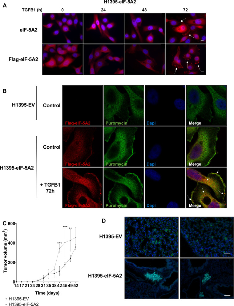
Overexpression of eIF-5A2 colocalizes with puromycin and promotes tumor metastasis in vivo. A Representative fluorescence microscopy images of H1395-eIF-5A2 cells untreated or treated with TGFB1 for the indicated times. Cells were stained with anti-eIF-5A2 or anti-Flag antibodies, and Dapi (blue) to observe the nuclei. Scale bar 10 µm. B Visualisation of the translational activity of eIF-5A2. H1395-EV and and H1395-eIF-5A2 treated with puromycin in the presence or absence of TGFB ligand for 72 h were stained with anti-Flag (red) and anti-Puromycin (green) antibody to visualize active places of translation in cells. Dapi (blue) staining was used to visualize nuclei. Scale bar 10 µm. C Overexpression of eIF-5A2 promotes tumor metastasis in vivo. H1395-EV and H1395-eIF-5A2 cells were subcutaneously injected in NSG mice. Tumor volume in the subcutaneous xenograft model was measured every 3–4 days while the condition of the mice was checked. Approximately two months after implantation, mice were euthanized and dissected (left panel). D Representative fluorescence microscopy images of lung metastasis of NSG mice injected as described in C. H1395-EV and H1395-eIF-5A2 metastasic cells expressing GFP is shown in green. Tissue was stained with Dapi (blue) to visualize nuclei. Scale bar for H1395-EV cells images 50 µm; scale bar for H1395-eIF-5A2 cells images 100 µm)

