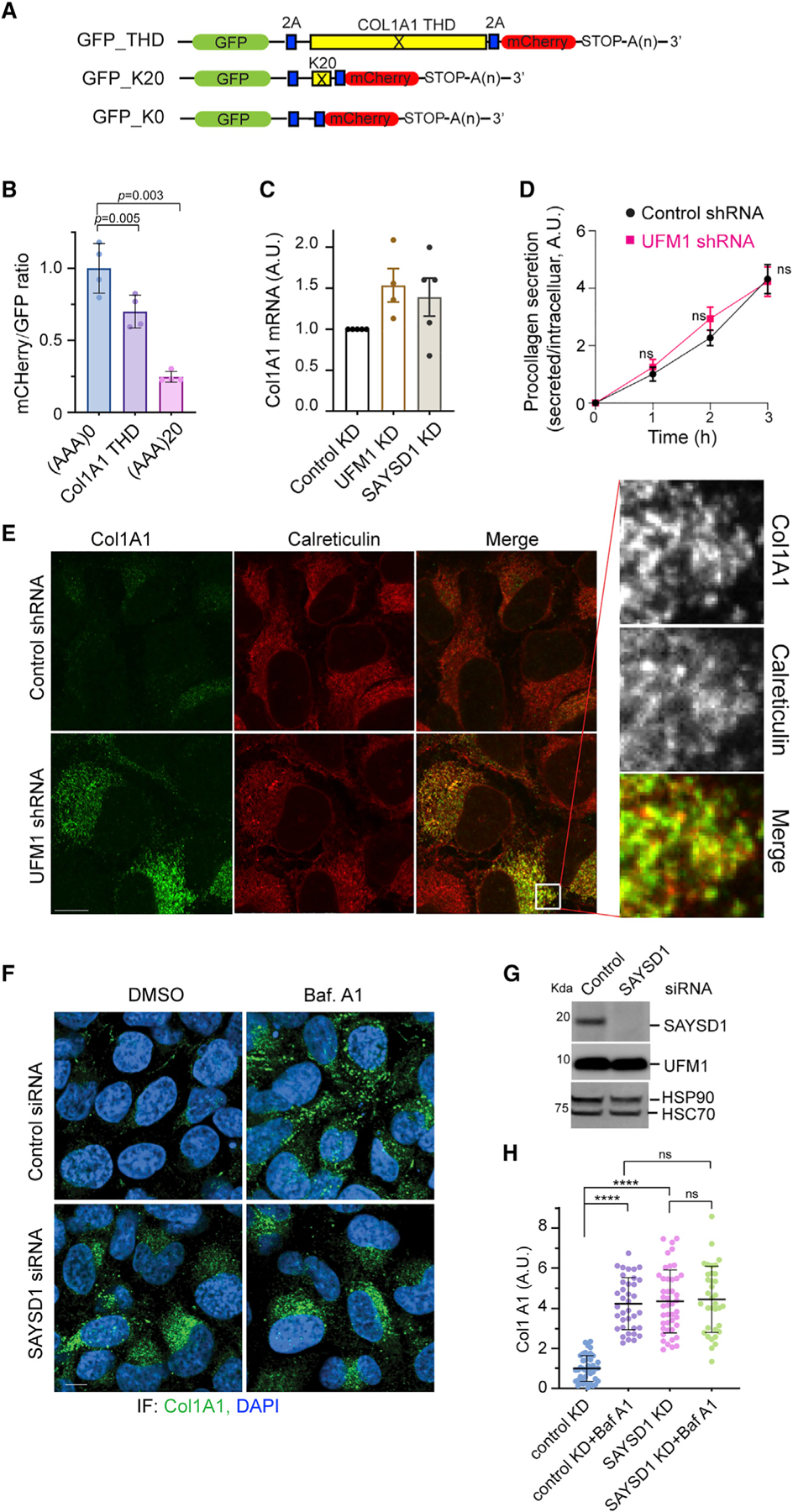
Figure 5. UFM1- and SAYSD1-dependent TAQC degrades translation-stalled Col1A1.

(A) A schematic view of the translation stalling reporters and the K0 control.
(B) The translation of the Col1A1 triple-helical domain (THD) causes ribosome stalling. 293T cells were transfected with the indicated reporters. The ratio of mC versus GFP was determined by flow cytometry. Error bars, means ± SD; p values are determined by one-way ANOVA followed by Dunnett’s multiple comparisons test; n = 4 independent experiments.
(C) UFM1 or SAYSD1 knockdown does not significantly increase Col1A1 mRNA as determined by qRT-PCR. n = 3 independent experiments. Error bars, means ± SD.
(D) UFM1 depletion does not affect Col1A1 secretion. Conditioned medium from control or UFM1-depleted U2OS cells were analyzed by ELISA. Error bars indicate means ± SD. ns, not significant by unpaired Student’s t test, n = 3 independent experiments.
(E) UFM1 depletion stabilizes Col1A1 in the ER. Control and UFM1 knockdown U2OS cells were stained by Col1A1 (green) and calreticulin (red) antibodies. Scale bar, 10 μm. Right panels show an enlarged view of the boxed area.
(F–H) Baf A1 treatment does not further enhance SAYSD1-depletion-induced Col1A1 accumulation.
(F) Control and SAYSD1 knockdown U2OS cells were treated with DMSO as a control or with Baf A1 (200 nM, 5 h) and stained by Col1A1 antibodies (green) or DAPI (blue). Scale bar, 10 μm. (G) A fraction of the siRNA-treated cells in (F) were analyzed by immunoblotting to confirm the knockdown of SAYSD1. (H) Quantification of the Col1A1 levels in (F). Error bars, means ± SD; ****p < 0.0001, by one-way ANOVA with Dunnett’s multiple comparisons test. n = 3 independent experiments.
