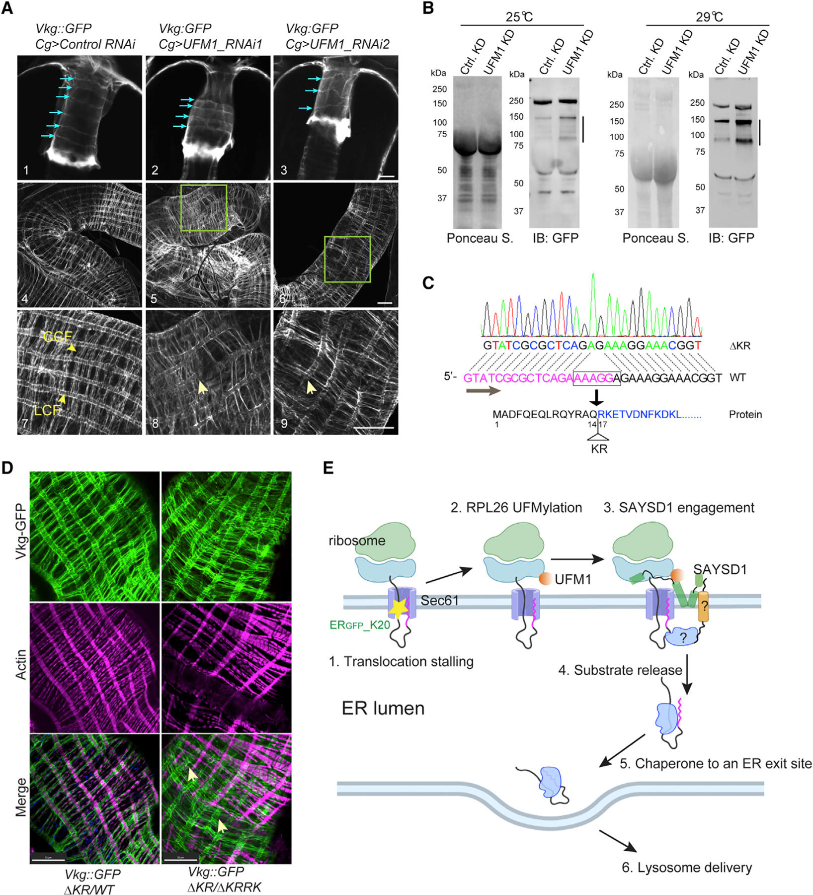
Figure 7. TAQC safeguards collagen biogenesis in Drosophila.

(A) Abnormal Viking deposition by UFM1-depleted FB in fly larvae. Shown are representative confocal images of Vkg-GFP on proventriculus (panels 1–3) or middle midgut (panels 4–9) from third-instar larvae of the indicated genotypes. Blue arrows in panels 1–3 indicate collagen fibrils around the proventriculus. Arrows in panels 8 and 9 indicate disrupted LCFs. Panels 7–9 are enlarged views. Scale bars, 20 μm.
(B) Defective Vkg-GFP accumulation in hemolymph in larvae with FB-specific UFM1 depletion. The lines indicate truncated Vkg-GFP species.
(C) Identification of a second SAYSD1 allele deleting two charged residues in the N17 domain.
(D) Abnormal Viking-containing basement membranes on middle midgut of fly larvae bearing the indicated mutant SAYSD1 alleles. The guts from third-instar larvae were stained by phalloidin to label actin (magenta). Arrows indicate Vkg-GFP-containing fibrils that are detached from muscle cells. Scale bars, 50 μm.
(E) A model of ribosome UFMylation sensing by SAYSD1 in TAQC. Created by Biorender.com.
