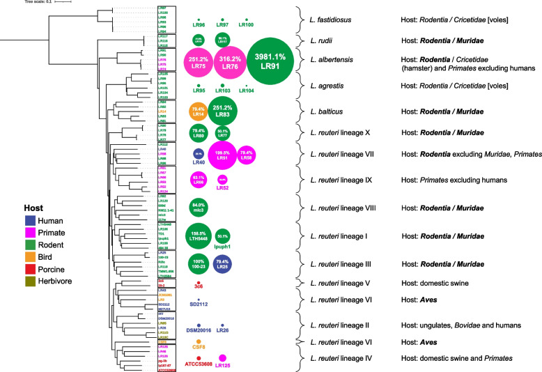
Fig. 7.
Colonization of the forestomach of germ-free mice by L. reuteri and related Limosilactobacillus species. The ML phylogenetic tree was constructed based the core-genome alignment of the representative strains from each of ten L. reuteri lineages and five Limosilactobacillus species. Adherent cell numbers of strains are represented by the area of the bubble besides the tree. The area of the bubble is scaled to be proportional to the log transformed cell counts that are expressed as % relative to strain L. reuteri subsp. rodentium 100–23. The cell counts are also shown in Additional file 4: Table S4 (L. reuteri strains) and Additional file 4: Table S5 (other five Limosilactobacillus species). Cell densities of more than 6.9 log10 of CFU/g, 20% relative to L. reuteri subsp. rodentium 100–23, were considered as effective epithelial adhesion; cell densities of less than 10%, corresponding to less than 6.6 log10 of CFU/g, were considered as ineffective epithelial adhesion. Shown to the right are the sources of isolation of strains of the respective linages. Taxonomic ranks of the respective hosts are printed in bold if host adaptation was demonstrated experimentally in this study or previously [17, 18, 39]

