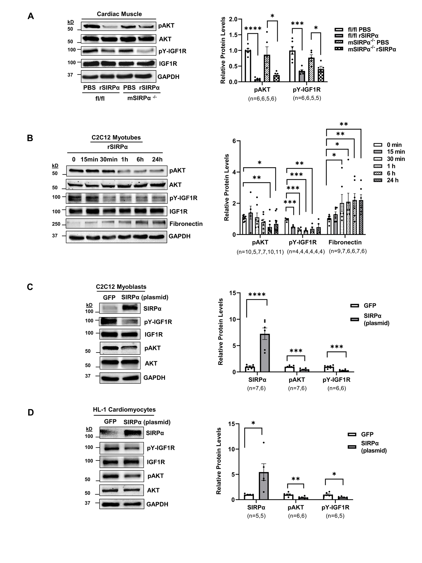
Figure 6. Both exogenous and intracellular SIRPα exacerbates insulin/IGF1 receptor responses.

(A) Flox (SIRPαfl/fl) and skeletal muscle-specific SIRPα KO (mSIRPα−/−) mice were treated with recombinant SIRPα protein (rSIRPα, 1 µg/g) or with diluent PBS control via left ventricular injection which was allowed to circulate for 5 min. Protein lysates of cardiac muscle were immunoblotted and representative immunoblots of averaged data (left panel) to detect pAKT relative to AKT and pY-IGF1R relative to IGF1R with densities (right panel) are shown. (B) C2C12 myotubes were treated with rSIRPα (1 µg/mL) and cell lysates were immunoblotted to detect pAKT relative to AKT, fibronectin, relative to GAPDH and pY-IGF1R relative to IGF1R and representative immunoblots of averaged data (left panel) are shown. The relative protein densities are shown (right panel). Myocytes (C) C2C12 myoblasts and (D) HL-1 cardiomyocytes were electroporated with 1.5 μg of GFP or 1.5 μg of SIRPα plasmid, protein lysates from cells were immunoblotted to detect SIRPα relative to GAPDH, pAKT relative to AKT, pY-IGF1R relative to IGF1R and representative immunoblots of averaged data (left panel) with relative densities (right panel) are shown. Statistical significance was calculated using unpaired two-tailed Studen's t-test (A-D). Values are means ± SEM. *p<0.05, ** p<0.01, *** p<0.001, **** p<0.0001.
