
Fig. 1.
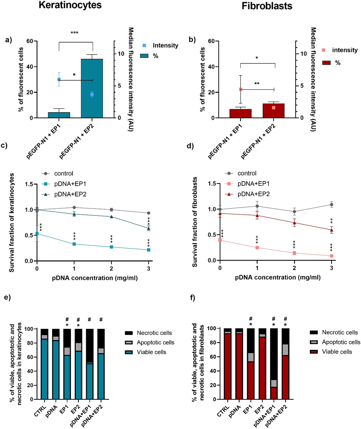
Transfection efficiency, cytotoxicity, and cell death mechanisms after electrotransfer of pDNA to keratinocytes and fibroblasts. Transfection efficiency was measured by flow cytometry in a) keratinocytes and b) fibroblasts (n = 3). Cell survival was measured by metabolic activity in c) keratinocytes and d) fibroblasts (n = 3–4) data were normalized to control. Apoptosis and necrosis were quantified by detecting Annexin V and 7-AAD staining using flow cytometry in e) keratinocytes and f) fibroblasts (n = 3) data were normalized to control. In a and b * **p < 0.001, * *p < 0.01, *p < 0.05 compared to different pulse protocols; in c and d * **p < 0.001, * *p < 0.01, *p < 0.05 compared to control; in e and f *p < 0.05 refers to apoptotic cells, #p < 0.05 refers to necrotic cells.