
Fig. 1.
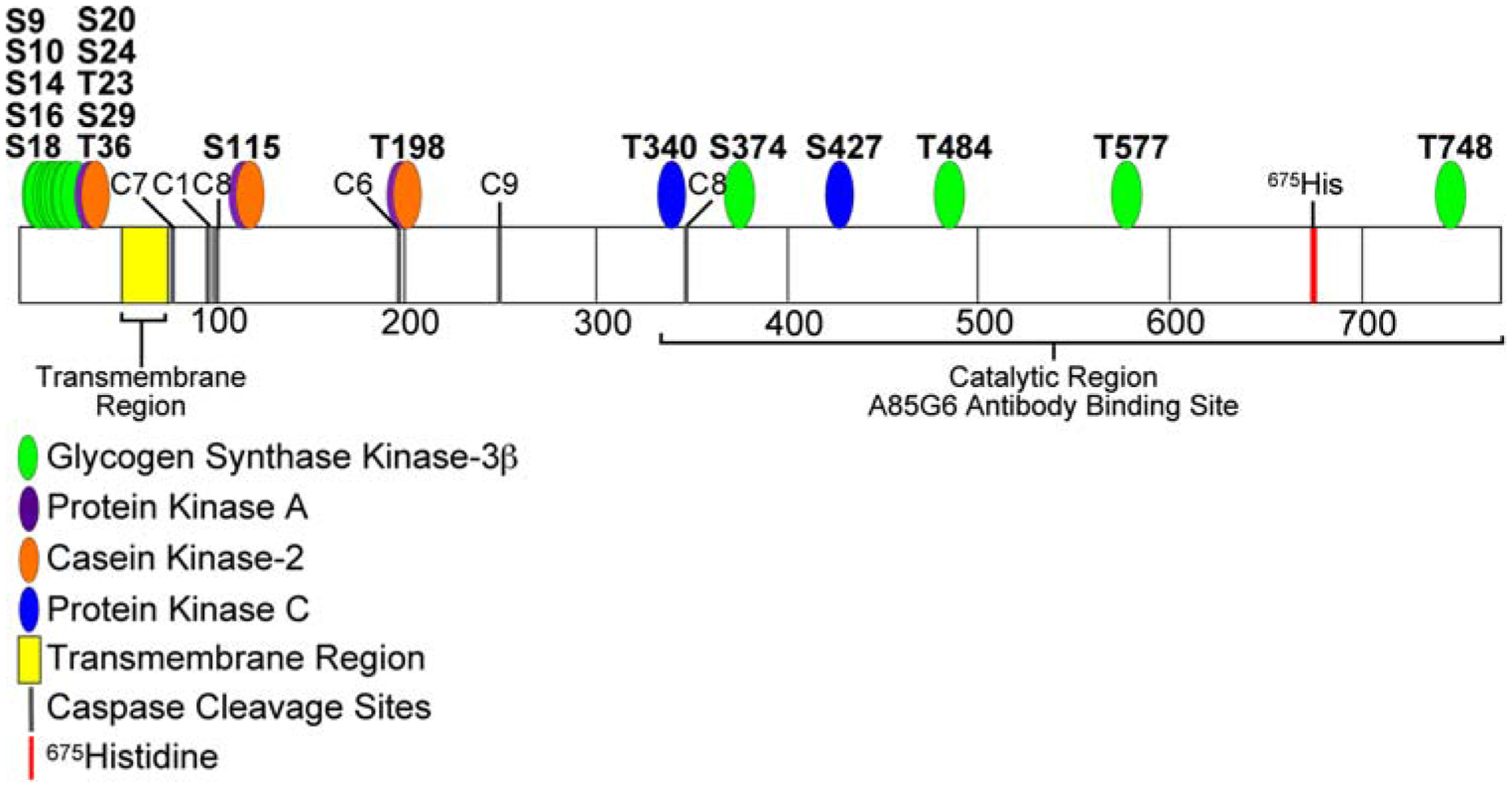
Diagram of predicted AAH phosphorylation sites. The translated amino acid sequence of human AAH was analyzed using the Long Peptide Subsequence Analysis module of MacVector 8.1 software. Positions of amino acids 100, 200, 300, 400, 500, 600, and 700 are indicated. Disks overlap with predicted Ser or Thr phosphorylation sites for glycogen synthase kinase 3β (GSK-3β-green), protein kinase A (PKA-violet), casein kinase 2 (CK II-orange), or protein kinase C (PKC-blue) phosphorylation. Note that the majority of the predicted sites (N = 13 of 24) have a consensus sequence corresponding to GSK-3β phosphorylation and are located within the N-terminal region of AAH protein. The transmembrane region (yellow), position of 675His (red), which is critical for AAH’s catalytic activity, and the C-terminal large cleavage fragment that has catalytic activity and is distinct from Humbug (underlined) are indicated