
Fig. 5.
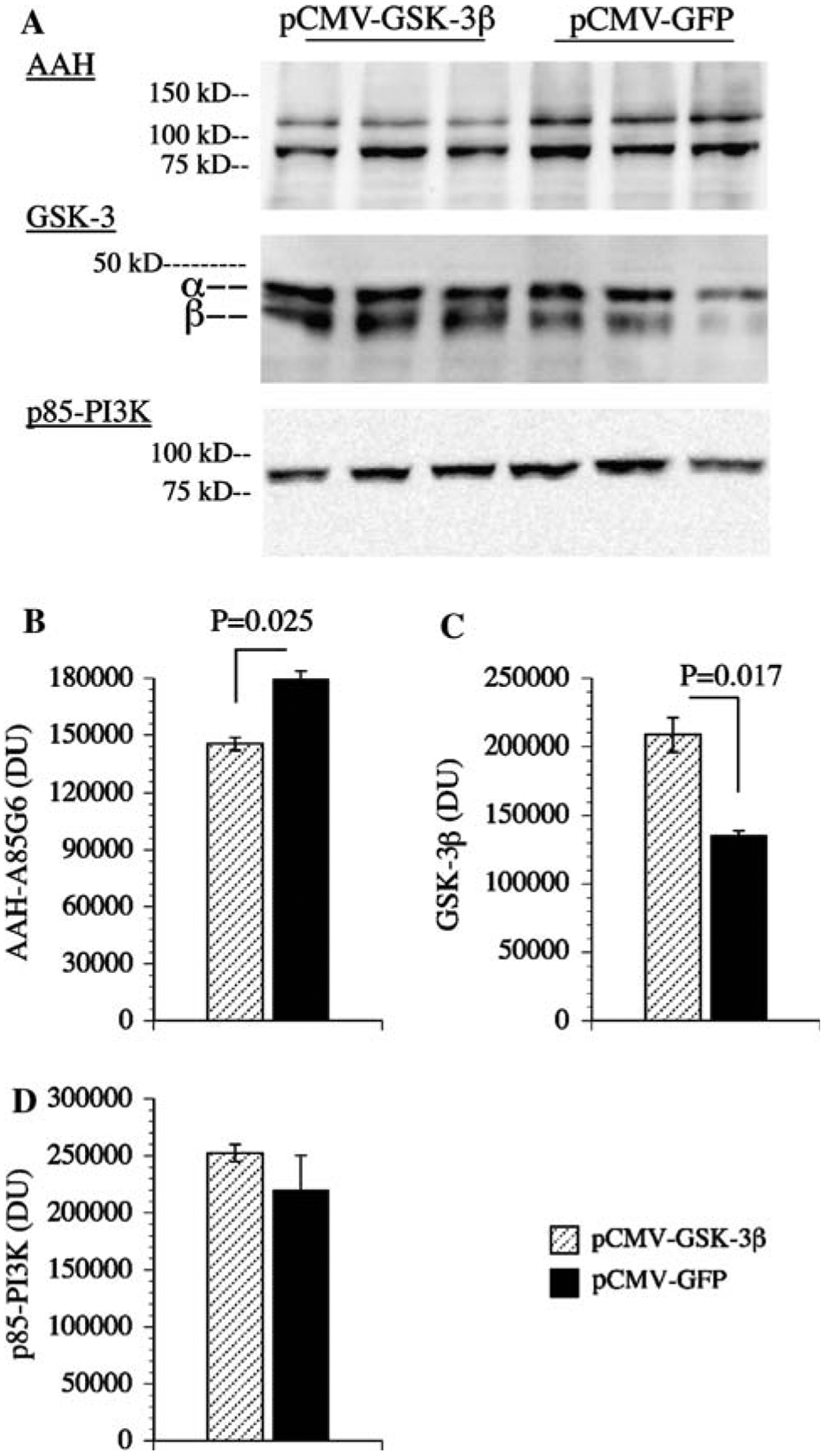
Overexpression of GSK-3β reduces AAH protein expression: PNET2 cells were transfected with recombinant plasmid containing GSK-3β (pGSKβ) or green fluorescent protein (pGFP) cDNA using Amaxa electroporation. a 48 h after transfection, cells were examined for AAH, GSK-3β, and p85 (PI3K-negative control) immunoreactivity by Western blot analysis. Levels of b AAH, c GSK-3, and d p85 immunoreactivities were quantified by digital imaging of the Western blot signals. Graphs depict mean ± SEM levels of immunoreactivity. Inter-group statistical comparisons were made using Student t-tests and significant P-values are indicated over the bars