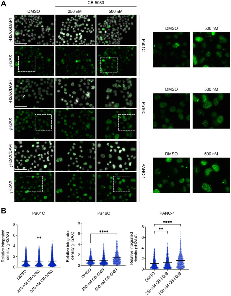
Figure 4. VCP, a regulator of DDR, helps mediate DNA damage repair in PDAC.
(A) Representative images of immunofluorescence (IF) to monitor γH2AX expression (green) and nuclei (white) in PDAC cells after 24 hours of treatment with DMSO or CB-5083 at the indicated concentrations (nM). Scale bar, 75 μm. (B) Quantitation of relative integrated intensity of γH2AX per nucleus of cells shown in panel A. Each dot represents a nucleus; each bar indicates the mean of that treatment group. Statistical significance was calculated using one-way ANOVA and Kruskal-Wallis test relative to DMSO. **p < 0.01 and ****p < 0.0001. The total number of nuclei analyzed for each cell line and condition (DMSO, 250 nM CB-5083, 500 nM CB-5083) were: Pa01C (1460, 1143, 1495), Pa16C (545, 746, 592), and PANC-1 (962, 902, 588), respectively.

