. 2023 Mar 13;12:e75906. doi: 10.7554/eLife.75906

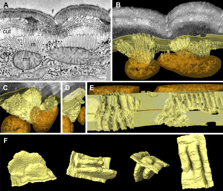
Figure 2. Meisosomes are formed by epidermal plasma membrane facing the apical extracellular matrix (ECM).

Adjacent meisosomes in two serial thick (300 nm) sections were analysed with by electron tomography. (A) Selected virtual image from the serial reconstruction. (B–E) Segmentation of membranes and mitochondria reveal their 3D topology. Meisosomes (in yellow) are in close apposition to, but not in continuity with, mitochondria (orange) and are formed by epidermal plasma membrane folds, as observed in an en face view after removing the cuticle in silico (E). (F) Two folds were extracted and manually filled for a schematic view. Cuticle (cut), epidermis (epi), furrow (f), mitochondria (mit), and meisosomes (m); scale bar, 200 nm.

Figure 2.

Figure 2—figure supplement 1. Meisosome distribution by serial block-face scanning electron microscopy (SBF-SEM).
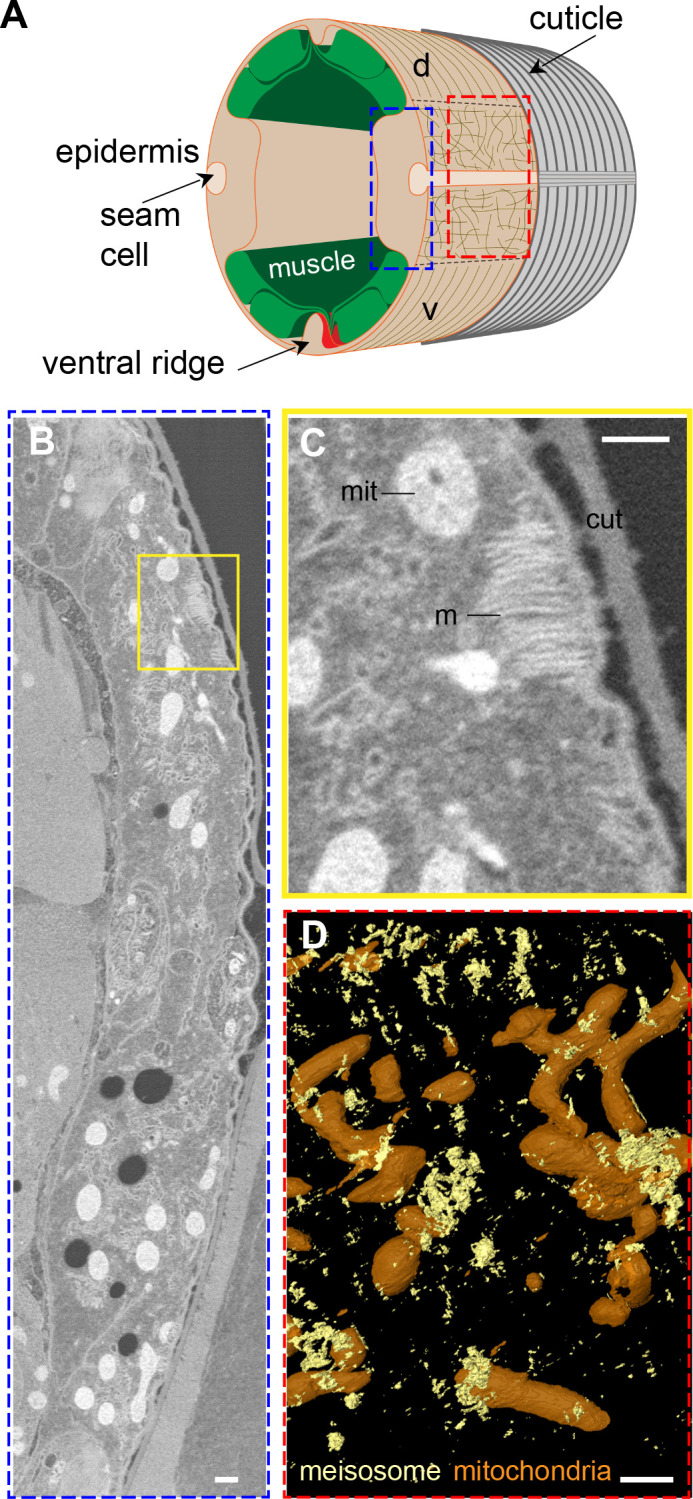
Figure 2—figure supplement 1.

(A) Schematic view of the outer tissues of an adult C. elegans worm near the mid-body. The plane of transverse EM sections and the orthogonal en face view for EM and fluorescence microscopy are indicated with the blue and red dashed rectangles, respectively. (B, C) Representative image from a serial block-face scanning electron microscopy (SBF-SEM) acquisition, the boxed area in (B) is shown at a higher magnification in (C). Scale bar, 500 nm. (D) SBF segmentation and 3D reconstruction revealed the en face spatial distribution of meisosomes (yellow) compared to mitochondria (orange). Scale bar, 1 µm.
Figure 2—video 1. Visualisation of the electron tomography and the 3D segmentation of meisosomes.
Download video file (20.2MB, mp4)
Two serial tomograms were stitched and segmentations of the plasma membrane, mitochondria, and cuticle were made (see ‘Materials and methods’ for details). The continuity of the plasma membrane could be revealed by semi-automated segmentation, except under certain orientation due the high membrane curvature of the meisosomes and the missing wedge artefact, which is inherent to electron tomography. At the end of the video, the segmentation of two folds was completed manually to present a schematic view.