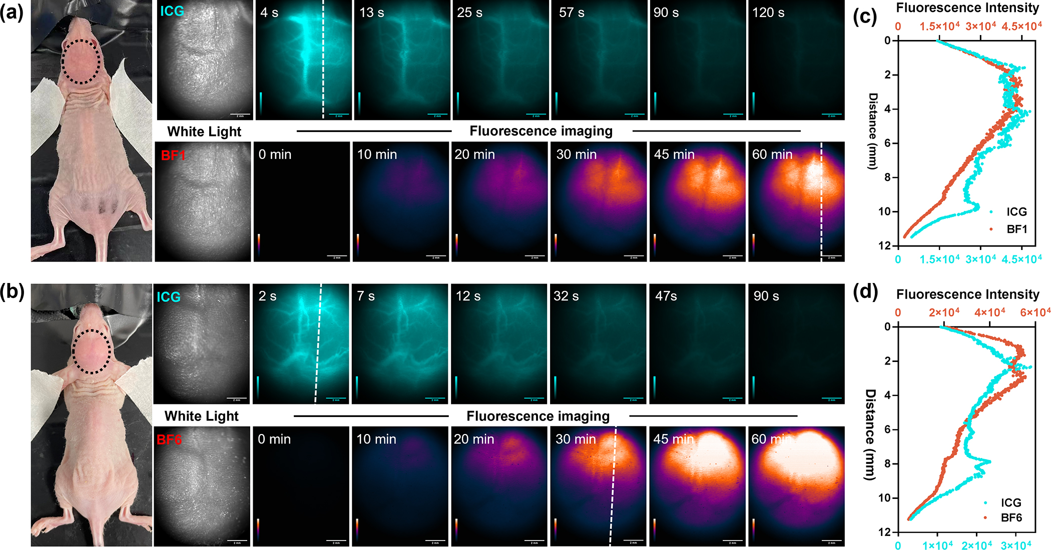
Figure 4.

(a) White light and noninvasively in vivo NIR-II fluorescence imaging of mouse brains bearing primary glioma tumor after treating ICG and BF1 (200 μM, 200 uL, tail vein injection) at six time points (4, 13, 25, 57, 90, 120 s for ICG; 0, 10, 20, 30, 45, 60 min for BF1). (b) White light and noninvasively in vivo NIR-II fluorescence imaging of mouse brains bearing primary glioma tumor after treating ICG and BF6 (200 μM, 200 uL, tail vein injection) at six time points (2, 7, 12, 32, 47, 90 s for ICG; 0, 10, 20, 30, 45, 60 min for BF6), respectively. The laser power was 70 mW cm−2, 808 nm laser, 1000 nm LP filter; (c, d) Cross-sectional fluorescence intensity profile along the white dash lines in (a) and (b) collected by 1000 nm long-pass filter.
