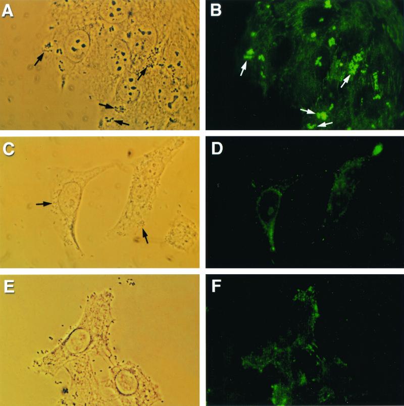
FIG. 1.
Reduced A/E lesion formation with phospholipase C inhibitor ET-18-OCH3 (approximate magnification, ×1,250). (A) Phase-contrast micrograph demonstrating adherent EPEC strain E2348/69 (arrows) on HEp-2 monolayers after coincubation for 3 h at 37°C. (B) Corresponding fluorescence micrograph showing α-actinin accumulation, demonstrated by bright foci of fluorescence (arrows), underneath adherent microcolonies of bacteria. (C) Phase-contrast micrograph showing adherent E2348/69 on ET-18-OCH3-pretreated epithelial cells (arrows). (D) α-Actinin accumulation under adherent EPEC was not observed in the corresponding fluorescence micrograph. (E) Phase-contrast micrograph demonstrating UMD864 adherent to tissue culture cells. (F) A negative α-actinin response was detected when HEp-2 cells were infected with a signaling-deficient mutant.

