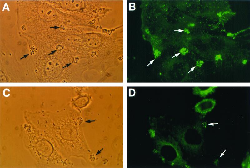
FIG. 2.
Reduced A/E lesion formation with phosphoinositide 3-kinase inhibitor wortmannin (10 nM) (approximate magnification, ×1,250). (A) EPEC strain E2348/69 showing initial adherence to tissue culture HEp-2 cells by phase-contrast microscopy (arrows). (B) Intense foci of fluorescence demonstrate reaggregation of α-actinin (arrows) corresponding to areas of bacterial attachment. (C) Phase-contrast micrograph depicting adherent E2348/69 on wortmannin-pretreated HEp-2 cells (arrows). (D) Fewer foci of α-actinin accumulation (arrows) under attaching bacteria were detected in the corresponding fluorescence micrograph.

