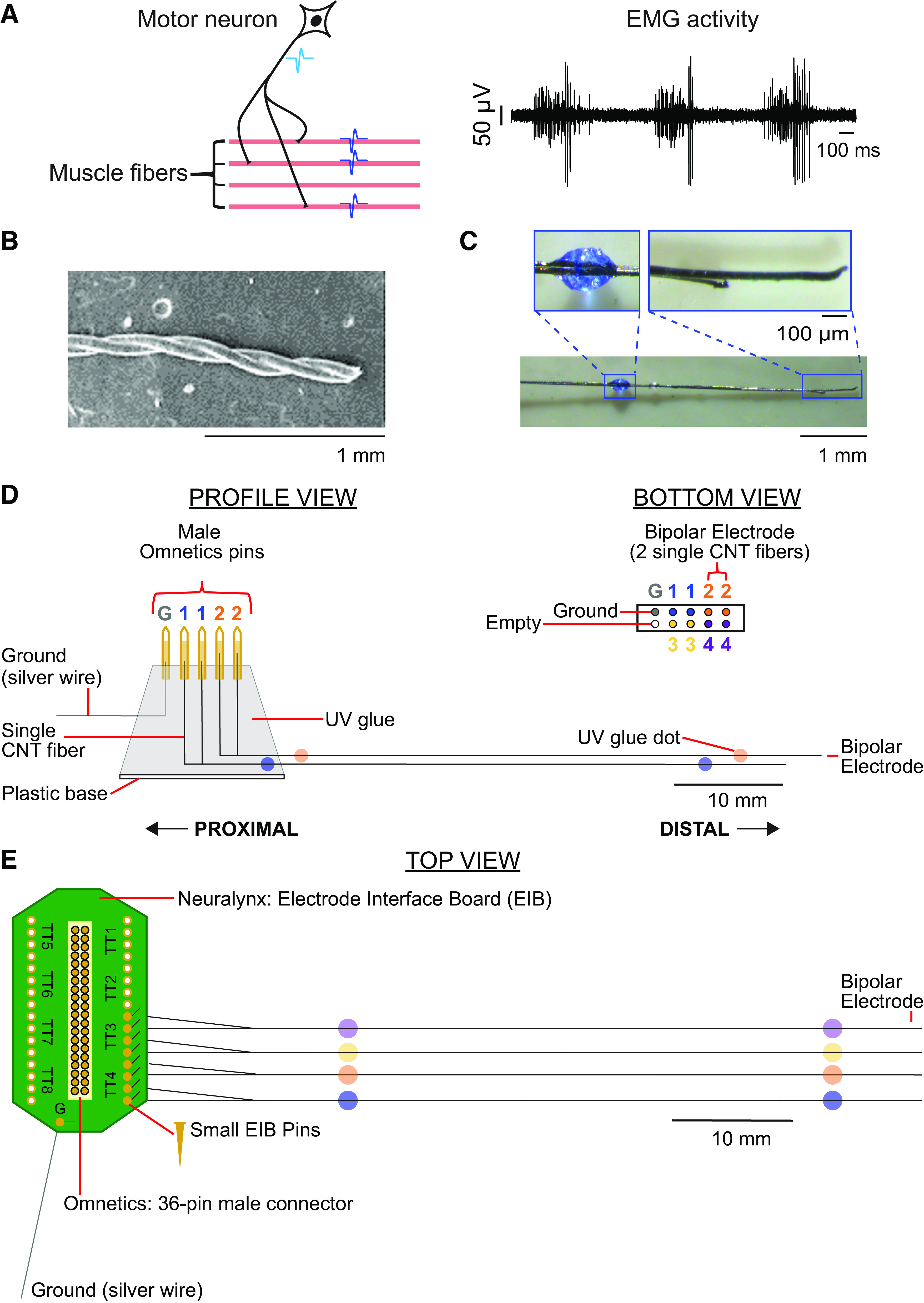
Figure 1.

Motor units, EMG, and carbon nanotube fibers (CNTFs) array construction. A, left: schematic of a motor unit, which consists of all muscle fibers (shown in pink) innervated by a single-motor neuron (shown in black). When a motor neuron fires an action potential (light blue waveform), the action potential moves down the axon to the muscle fibers it innervates, causing the innervated muscle fibers to fire a near-synchronous volley of action potentials (dark blue waveforms). Right: example recordings of multiunit EMG activity. B: scanning electron microscopy image of two parylene-coated carbon nanotube fibers twisted together, creating a bipolar electrode. C: each bipolar electrode was labeled with colored dots of ultraviolet (UV) glue. Each electrode array consisted of four CNTF bipolar electrodes and one ground wire. D: CNTF array schematic for design 1. Each proximal end of four CNTF bipolar electrodes and one ground wire were secured into male Omnetics pins with carbon glue, creating a two by five electrode array. E: CNTF array schematic for design 2. Each proximal end of a CNTF bipolar electrode was inserted into side-by-side gold-plated holes on a Neuralynx electrode board interface and secured with a gold attachment pin.
